Altintoprak Fatih, Degirmenci Bumin, Dikicier Enis, Cakmak Guner, Kivilcim Taner, Akbulut Gokhan, Dilek Osman Nuri, Gunduz Yasemin
Department of General Surgery, Faculty of Medicine, Sakarya University, Turkey.
ScientificWorldJournal. 2013 Apr 18;2013:298392. doi: 10.1155/2013/298392. Print 2013.
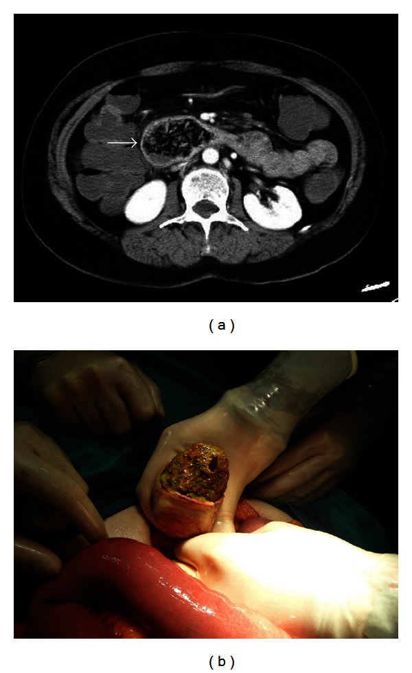
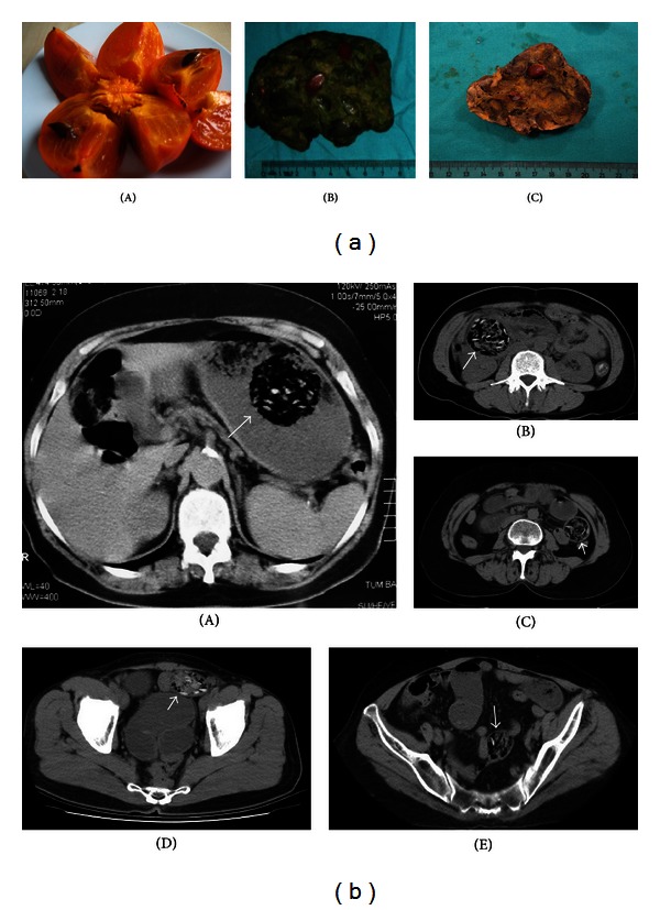
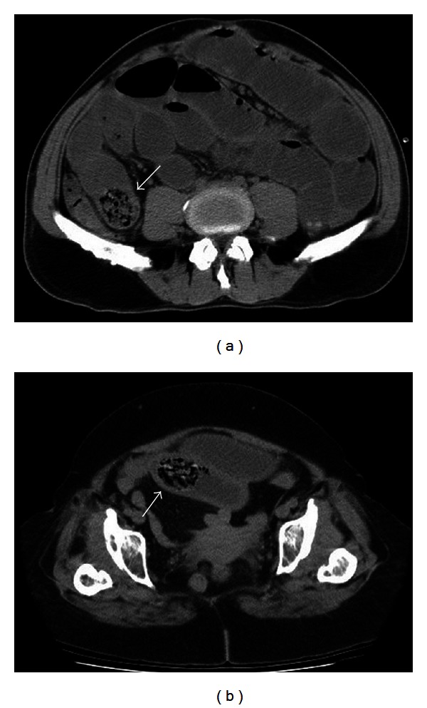
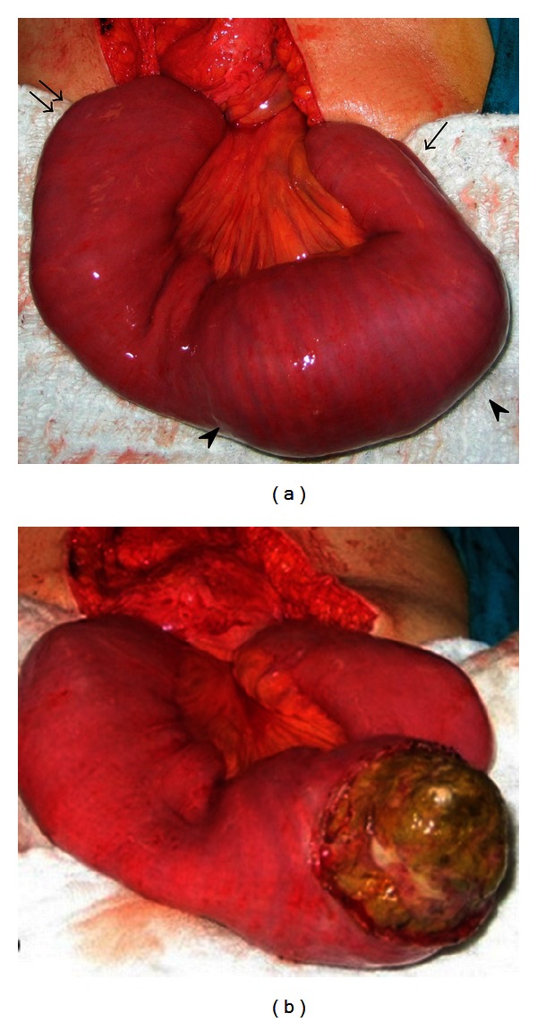
PURPOSE. The aim of this study was to present the computed tomography (CT) findings of bezoars that cause obstruction in the small bowel and to emphasize that some CT findings can be considered specific to some bezoar types. MATERIALS AND METHODS. The records of 39 patients who underwent preoperative abdominal CT and subsequent operation with a diagnosis of intestinal obstruction due to bezoars were retrospectively analyzed. RESULTS. In total, 56 bezoars were surgically removed from 39 patients. Bezoars were most commonly located in the jejunum (n = 26/56, 46.4%). Sixteen (41.0%) patients had multiple bezoar locations in the gastrointestinal tract. Common CT findings in all patients were a mottled gas pattern and a focal ovoid or round intraluminal mass with regular margins and a heterogeneous internal structure. Furthermore, some CT findings were determined to be specific to bezoars caused by persimmons. CONCLUSIONS. Preoperative CT is valuable in patients admitted with signs of intestinal obstruction in geographic regions with a high bezoar prevalence. We believe that the correct diagnosis of bezoars and the identification of their number and location provide a great advantage for all physicians and surgeons. In addition, some types of bezoars have unique CT findings, and we believe that these findings may help to establish a diagnosis.
目的。本研究的目的是呈现导致小肠梗阻的胃石的计算机断层扫描(CT)表现,并强调某些CT表现可能被认为是某些类型胃石所特有的。材料与方法。回顾性分析39例因胃石导致肠梗阻而接受术前腹部CT检查及后续手术的患者的记录。结果。39例患者共手术取出56枚胃石。胃石最常见于空肠(n = 26/56,46.4%)。16例(41.0%)患者胃肠道有多个胃石位置。所有患者的常见CT表现为斑片状气体影和腔内局灶性卵圆形或圆形肿块,边缘规则,内部结构不均匀。此外,一些CT表现被确定为柿子所致胃石所特有。结论。在胃石患病率高的地区,术前CT对肠梗阻征象患者有价值。我们认为,正确诊断胃石并确定其数量和位置对所有内科医生和外科医生都有很大优势。此外,某些类型的胃石有独特的CT表现,我们认为这些表现可能有助于确诊。