Schroeder Kristopher M, Ramamoorthy Jagan, Galgon Richard E
Department of Anesthesiology, University of Wisconsin School of Medicine and Public Health, 600 Highland Avenue, B6/319 CSC, Madison, WI, USA.
Indian J Anaesth. 2013 Jan;57(1):31-4. doi: 10.4103/0019-5049.108558.
Recent manuscripts have described the use of ultrasound imaging to evaluate airway structures. Ultrasound training tools are necessary for practitioners to become proficient at obtaining and interpreting images. Few training tools exist and those that do can often times be expensive and rendered useless with repeated needle passes.
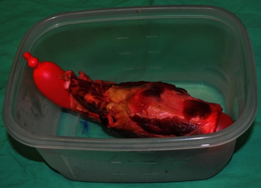
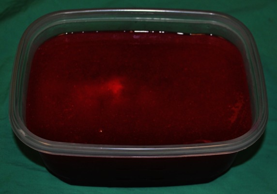
We utilised inexpensive and easy to obtain materials to create a gel phantom model for ultrasound-guided airway examination training.
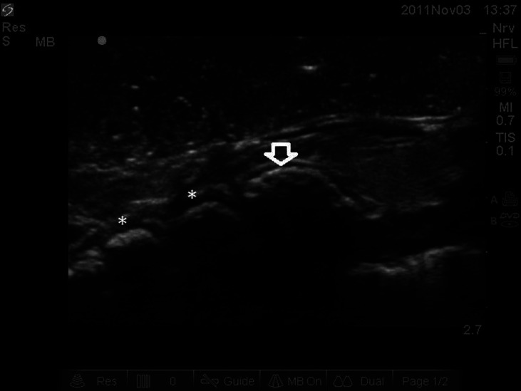
Following creation of the gel phantom model, images were successfully obtained of the thyroid and cricoid cartilages, cricothyroid membrane and tracheal rings in both the sagittal transverse planes.
The gel phantom model mimics human airway anatomy and may be used for ultrasound-guided airway assessment and intervention training. This may have important safety implications as ultrasound imaging is increasingly used for airway assessment.
近期的文献描述了使用超声成像来评估气道结构。超声训练工具对于从业者熟练获取和解读图像是必要的。现有的训练工具很少,而且那些工具往往很昂贵,并且在反复进行穿刺后会变得无用。
我们利用廉价且易于获取的材料制作了一个用于超声引导气道检查训练的凝胶体模模型。
在制作凝胶体模模型后,成功在矢状面和横断面上获取了甲状腺、环状软骨、环甲膜和气管环的图像。
凝胶体模模型模拟了人体气道解剖结构,可用于超声引导气道评估和干预训练。随着超声成像越来越多地用于气道评估,这可能具有重要的安全意义。