• 文献检索
  • 文档翻译
  • 深度研究
  • 学术资讯
  • Suppr Zotero 插件Zotero 插件
  • 邀请有礼
  • 套餐&价格
  • 历史记录
应用&插件
Suppr Zotero 插件Zotero 插件浏览器插件Mac 客户端Windows 客户端微信小程序
定价
高级版会员购买积分包购买API积分包
服务
文献检索文档翻译深度研究API 文档MCP 服务
关于我们
关于 Suppr公司介绍联系我们用户协议隐私条款
关注我们

Suppr 超能文献

核心技术专利:CN118964589B侵权必究
粤ICP备2023148730 号-1Suppr @ 2026

文献检索

告别复杂PubMed语法,用中文像聊天一样搜索,搜遍4000万医学文献。AI智能推荐,让科研检索更轻松。

立即免费搜索

文件翻译

保留排版,准确专业,支持PDF/Word/PPT等文件格式,支持 12+语言互译。

免费翻译文档

深度研究

AI帮你快速写综述,25分钟生成高质量综述,智能提取关键信息,辅助科研写作。

立即免费体验

脑创伤后 MRI 和组织学测量的神经突密度比较。

Comparison of neurite density measured by MRI and histology after TBI.

机构信息

Department of Neurology, Henry Ford Hospital, Detroit, Michigan, USA.

出版信息

PLoS One. 2013 May 22;8(5):e63511. doi: 10.1371/journal.pone.0063511. Print 2013.

DOI:10.1371/journal.pone.0063511
PMID:23717439
原文链接:https://pmc.ncbi.nlm.nih.gov/articles/PMC3661595/
Abstract

BACKGROUND

Functional recovery after brain injury in animals is improved by marrow stromal cells (MSC) which stimulate neurite reorganization. However, MRI measurement of neurite density changes after injury has not been performed. In this study, we investigate the feasibility of MRI measurement of neurite density in an animal model of traumatic brain injury (TBI) with and without MSC treatment.

METHODS

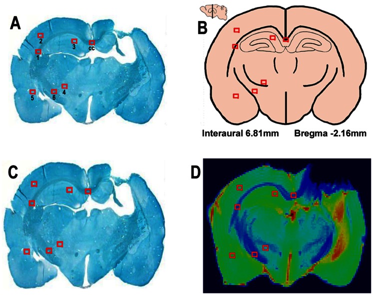
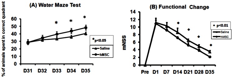
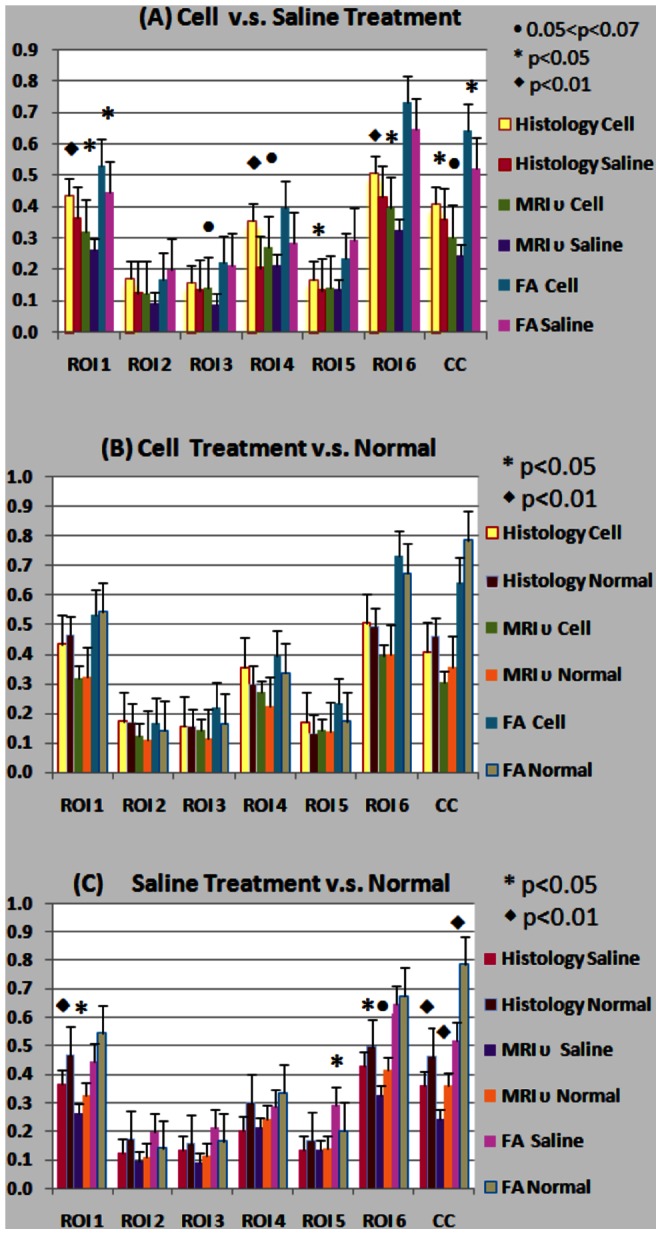
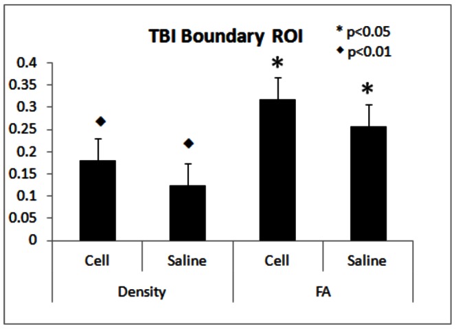
Fifteen male Wistar rats, were treated with saline (n = 6) or MSCs (n = 9) and were sacrificed at 6 weeks after controlled cortical impact (CCI). Healthy non-CCI rats (n = 5), were also employed. Ex-vivo MRI scans were performed two days after the rats were sacrificed. Multiple-shell hybrid diffusion imaging encoding scheme and spherical harmonic expansion of a two-compartment water diffusion displacement model were used to extract neurite related parameters. Bielshowski and Luxol Fast blue was used for staining axons and myelin, respectively. Modified Morris water maze and neurological severity score (mNSS) test were performed for functional evaluation. The treatment effects, the correlations between neurite densities measured by MRI and histology, and the correlations between MRI and functional variables were calculated by repeated measures analysis of variance, the regression correlation analysis tests, and spearman correlation coefficients.

RESULTS

Neurite densities exhibited a significant correlation (R(2)>0.80, p<1E-20) between MRI and immuno-histochemistry measurements with 95% lower bound of the intra-correlation coefficient (ICC) as 0.86. The conventional fractional anisotropy (FA) correlated moderately with histological neurite density (R(2) = 0.59, P<1E-5) with 95% lower bound of ICC as 0.76. MRI data revealed increased neurite reorganization with MSC treatment compared with saline treatment, confirmed by histological data from the same animals. mNSS were significantly correlated with MRI neurite density in the hippocampus region.

CONCLUSIONS

The present studies demonstrated that neurite density can be estimated by MRI after TBI and MRI measurement of neurite density is a sensitive marker to MSC treatment response.

摘要

背景

骨髓基质细胞(MSC)可刺激轴突重组,从而改善脑损伤后的功能恢复。然而,尚未对损伤后轴突密度变化的 MRI 测量进行研究。在这项研究中,我们探讨了在创伤性脑损伤(TBI)动物模型中使用和不使用 MSC 治疗时,MRI 测量轴突密度的可行性。

方法

15 只雄性 Wistar 大鼠,分别用生理盐水(n=6)或 MSC(n=9)处理,并在皮质撞击(CCI)后 6 周处死。还使用了健康的非 CCI 大鼠(n=5)。大鼠死后两天进行离体 MRI 扫描。使用多壳混合扩散成像编码方案和双室水扩散位移模型的球谐展开来提取与轴突相关的参数。Bielshowski 和 Luxol Fast blue 分别用于轴突和髓鞘的染色。改良 Morris 水迷宫和神经功能严重程度评分(mNSS)测试用于功能评估。通过重复测量方差分析、回归相关分析测试和 Spearman 相关系数,计算治疗效果、MRI 和免疫组织化学测量的轴突密度之间的相关性,以及 MRI 和功能变量之间的相关性。

结果

MRI 与免疫组织化学测量之间的轴突密度呈显著相关性(R(2)>0.80,p<1E-20),内相关系数(ICC)的 95%下限为 0.86。常规各向异性分数(FA)与组织学轴突密度中度相关(R(2)=0.59,P<1E-5),ICC 的 95%下限为 0.76。与生理盐水治疗相比,MSC 治疗后 MRI 数据显示出更多的轴突重组,这与来自同一动物的组织学数据一致。mNSS 与海马区的 MRI 轴突密度显著相关。

结论

本研究表明,TBI 后可通过 MRI 估计轴突密度,MRI 测量的轴突密度是 MSC 治疗反应的敏感标志物。

https://cdn.ncbi.nlm.nih.gov/pmc/blobs/9ebb/3661595/e0b9ed47bc72/pone.0063511.g006.jpg
https://cdn.ncbi.nlm.nih.gov/pmc/blobs/9ebb/3661595/91b0dff56130/pone.0063511.g001.jpg
https://cdn.ncbi.nlm.nih.gov/pmc/blobs/9ebb/3661595/683c7b9d31a7/pone.0063511.g002.jpg
https://cdn.ncbi.nlm.nih.gov/pmc/blobs/9ebb/3661595/201d6be5866d/pone.0063511.g003.jpg
https://cdn.ncbi.nlm.nih.gov/pmc/blobs/9ebb/3661595/c6ac1e34d014/pone.0063511.g004.jpg
https://cdn.ncbi.nlm.nih.gov/pmc/blobs/9ebb/3661595/085dffb2965c/pone.0063511.g005.jpg
https://cdn.ncbi.nlm.nih.gov/pmc/blobs/9ebb/3661595/e0b9ed47bc72/pone.0063511.g006.jpg

相似文献

1
Comparison of neurite density measured by MRI and histology after TBI.脑创伤后 MRI 和组织学测量的神经突密度比较。
PLoS One. 2013 May 22;8(5):e63511. doi: 10.1371/journal.pone.0063511. Print 2013.
2
MRI evaluation of axonal reorganization after bone marrow stromal cell treatment of traumatic brain injury.MRI 评价骨髓基质细胞治疗创伤性脑损伤后轴突重排。
NMR Biomed. 2011 Nov;24(9):1119-28. doi: 10.1002/nbm.1667. Epub 2011 Mar 23.
3
MRI of neuronal recovery after low-dose methamphetamine treatment of traumatic brain injury in rats.MRI 观察低剂量冰毒治疗大鼠创伤性脑损伤后的神经元恢复。
PLoS One. 2013 Apr 18;8(4):e61241. doi: 10.1371/journal.pone.0061241. Print 2013.
4
Effects of treating traumatic brain injury with collagen scaffolds and human bone marrow stromal cells on sprouting of corticospinal tract axons into the denervated side of the spinal cord.胶原支架和人骨髓基质细胞治疗创伤性脑损伤对皮质脊髓束轴突向脊髓去神经侧发芽的影响。
J Neurosurg. 2013 Feb;118(2):381-9. doi: 10.3171/2012.11.JNS12753. Epub 2012 Nov 30.
5
Diffuse white matter response in trauma-injured brain to bone marrow stromal cell treatment detected by diffusional kurtosis imaging.弥散张量成像检测创伤性脑损伤后骨髓基质细胞治疗的弥散峰度反应。
Brain Res. 2019 Aug 15;1717:127-135. doi: 10.1016/j.brainres.2019.04.020. Epub 2019 Apr 19.
6
Marrow stromal cell transplantation after traumatic brain injury promotes cellular proliferation within the brain.创伤性脑损伤后进行骨髓基质细胞移植可促进脑内细胞增殖。
Neurosurgery. 2004 Nov;55(5):1185-93. doi: 10.1227/01.neu.0000141042.14476.3c.
7
Down-regulation of Nogo-A by collagen scaffolds impregnated with bone marrow stromal cell treatment after traumatic brain injury promotes axonal regeneration in rats.经骨髓基质细胞处理的胶原支架下调创伤性脑损伤后 Nogo-A 的表达,促进大鼠轴突再生。
Brain Res. 2014 Jan 13;1542:41-8. doi: 10.1016/j.brainres.2013.10.045. Epub 2013 Oct 28.
8
Characterizing brain structures and remodeling after TBI based on information content, diffusion entropy.基于信息内容和扩散熵描述 TBI 后的大脑结构和重塑。
PLoS One. 2013 Oct 15;8(10):e76343. doi: 10.1371/journal.pone.0076343. eCollection 2013.
9
Transplantation of marrow stromal cells restores cerebral blood flow and reduces cerebral atrophy in rats with traumatic brain injury: in vivo MRI study.骨髓基质细胞移植恢复创伤性脑损伤大鼠的脑血流并减少脑萎缩:体内 MRI 研究。
J Neurotrauma. 2011 Apr;28(4):535-45. doi: 10.1089/neu.2010.1619. Epub 2011 Mar 24.
10
Treatment of traumatic brain injury in mice with marrow stromal cells.用骨髓基质细胞治疗小鼠创伤性脑损伤。
Brain Res. 2008 May 7;1208:234-9. doi: 10.1016/j.brainres.2008.02.042. Epub 2008 Mar 4.

引用本文的文献

1
Volume electron microscopy in injured rat brain validates white matter microstructure metrics from diffusion MRI.对受伤大鼠大脑进行的体积电子显微镜检查验证了扩散磁共振成像中的白质微观结构指标。
Imaging Neurosci (Camb). 2024 Jul 2;2. doi: 10.1162/imag_a_00212. eCollection 2024.
2
Systematic review and meta-analysis of preclinical studies testing mesenchymal stromal cells for traumatic brain injury.对测试间充质基质细胞治疗创伤性脑损伤的临床前研究的系统评价和荟萃分析。
NPJ Regen Med. 2021 Oct 29;6(1):71. doi: 10.1038/s41536-021-00182-8.
3
From biomechanics to pathology: predicting axonal injury from patterns of strain after traumatic brain injury.

本文引用的文献

1
MRI detects brain reorganization after human umbilical tissue-derived cells (hUTC) treatment of stroke in rat.MRI 检测到人脐带组织来源的细胞(hUTC)治疗大鼠中风后脑组织重组。
PLoS One. 2012;7(8):e42845. doi: 10.1371/journal.pone.0042845. Epub 2012 Aug 10.
2
MRI evaluation of axonal reorganization after bone marrow stromal cell treatment of traumatic brain injury.MRI 评价骨髓基质细胞治疗创伤性脑损伤后轴突重排。
NMR Biomed. 2011 Nov;24(9):1119-28. doi: 10.1002/nbm.1667. Epub 2011 Mar 23.
3
Post-treatment with amphetamine enhances reinnervation of the ipsilateral side cortex in stroke rats.
从生物力学到病理学:从创伤性脑损伤后的应变模式预测轴突损伤。
Brain. 2021 Feb 12;144(1):70-91. doi: 10.1093/brain/awaa336.
4
Cellular and molecular imaging for stem cell tracking in neurological diseases.用于神经疾病中干细胞示踪的细胞和分子成像。
Stroke Vasc Neurol. 2021 Mar;6(1):121-127. doi: 10.1136/svn-2020-000408. Epub 2020 Oct 29.
5
Diffusion MR imaging acquisition and analytics for microstructural delineation in pre-clinical models of TBI.弥散磁共振成像采集和分析在创伤性脑损伤临床前模型中的微观结构描绘。
J Neurosci Res. 2022 May;100(5):1128-1139. doi: 10.1002/jnr.24416. Epub 2019 May 1.
6
Four in vivo g-ratio-weighted imaging methods: Comparability and repeatability at the group level.四种活体 g 比值加权成像方法:在组水平上的可比性和可重复性。
Hum Brain Mapp. 2018 Jan;39(1):24-41. doi: 10.1002/hbm.23858. Epub 2017 Nov 1.
7
Diffusion MRI and the detection of alterations following traumatic brain injury.弥散磁共振成像与颅脑创伤后改变的检测。
J Neurosci Res. 2018 Apr;96(4):612-625. doi: 10.1002/jnr.24065. Epub 2017 Jun 13.
8
Amide proton transfer-weighted MRI detection of traumatic brain injury in rats.酰胺质子转移加权磁共振成像检测大鼠创伤性脑损伤
J Cereb Blood Flow Metab. 2017 Oct;37(10):3422-3432. doi: 10.1177/0271678X17690165. Epub 2017 Jan 27.
9
Magnetic resonance imaging and cell-based neurorestorative therapy after brain injury.脑损伤后的磁共振成像与基于细胞的神经修复治疗
Neural Regen Res. 2016 Jan;11(1):7-14. doi: 10.4103/1673-5374.169603.
10
Application of neurite orientation dispersion and density imaging (NODDI) to a tau pathology model of Alzheimer's disease.神经突方向离散度与密度成像(NODDI)在阿尔茨海默病tau病理模型中的应用。
Neuroimage. 2016 Jan 15;125:739-744. doi: 10.1016/j.neuroimage.2015.10.043. Epub 2015 Oct 23.
阿朴吗啡治疗增强了卒中大鼠对侧皮质的神经再支配。
Neuroimage. 2011 May 1;56(1):280-9. doi: 10.1016/j.neuroimage.2011.02.049. Epub 2011 Feb 22.
4
Autologous bone marrow mononuclear cell therapy for severe traumatic brain injury in children.儿童严重创伤性脑损伤的自体骨髓单个核细胞治疗。
Neurosurgery. 2011 Mar;68(3):588-600. doi: 10.1227/NEU.0b013e318207734c.
5
Neuroprotection for traumatic brain injury: translational challenges and emerging therapeutic strategies.颅脑创伤的神经保护:转化医学的挑战与新兴治疗策略。
Trends Pharmacol Sci. 2010 Dec;31(12):596-604. doi: 10.1016/j.tips.2010.09.005. Epub 2010 Oct 29.
6
Orientationally invariant indices of axon diameter and density from diffusion MRI.基于弥散磁共振成像的各向同性轴突直径和密度指标
Neuroimage. 2010 Oct 1;52(4):1374-89. doi: 10.1016/j.neuroimage.2010.05.043. Epub 2010 May 23.
7
Exploring new routes for neuroprotective drug development in traumatic brain injury.探索创伤性脑损伤中神经保护药物开发的新途径。
Sci Transl Med. 2010 Apr 14;2(27):27rv1. doi: 10.1126/scitranslmed.3000330.
8
Neurite density from magnetic resonance diffusion measurements at ultrahigh field: comparison with light microscopy and electron microscopy.超高场磁共振扩散测量的神经突密度:与光学显微镜和电子显微镜的比较。
Neuroimage. 2010 Jan 1;49(1):205-16. doi: 10.1016/j.neuroimage.2009.08.053. Epub 2009 Sep 2.
9
Marrow stromal cell transplantation in stroke and traumatic brain injury.骨髓基质细胞移植治疗中风和创伤性脑损伤。
Neurosci Lett. 2009 Jun 12;456(3):120-3. doi: 10.1016/j.neulet.2008.03.096. Epub 2009 Jan 17.
10
Edaravone attenuates white matter lesions through endothelial protection in a rat chronic hypoperfusion model.依达拉奉通过保护内皮细胞减轻大鼠慢性脑灌注不足模型中的白质损伤。
Neuroscience. 2009 Aug 18;162(2):317-27. doi: 10.1016/j.neuroscience.2009.04.065. Epub 2009 May 3.