Department of Brain Sciences, Faculty of Medicine, Imperial College London, London, UK.
Royal British Legion Centre for Blast Injury Studies, Imperial College London, London, UK.
Brain. 2021 Feb 12;144(1):70-91. doi: 10.1093/brain/awaa336.
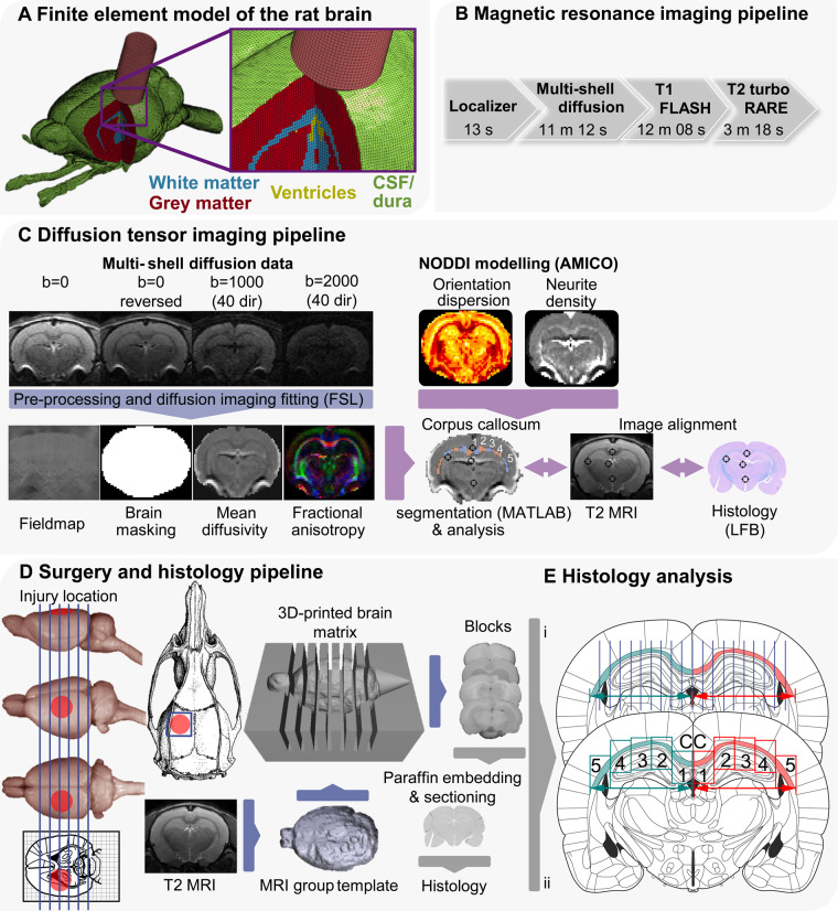
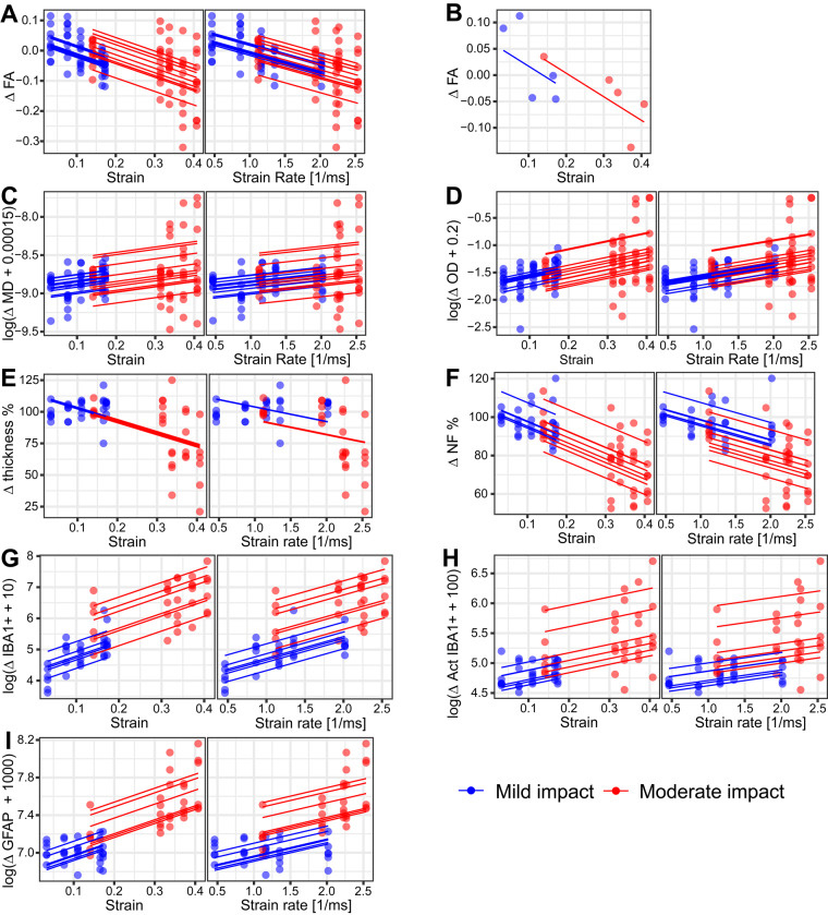
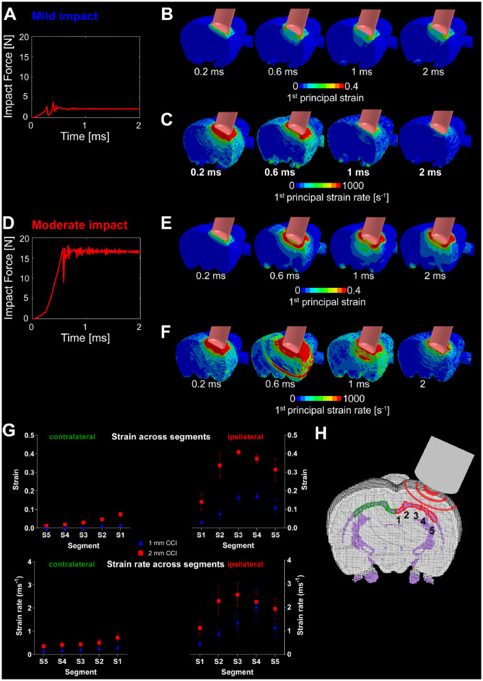
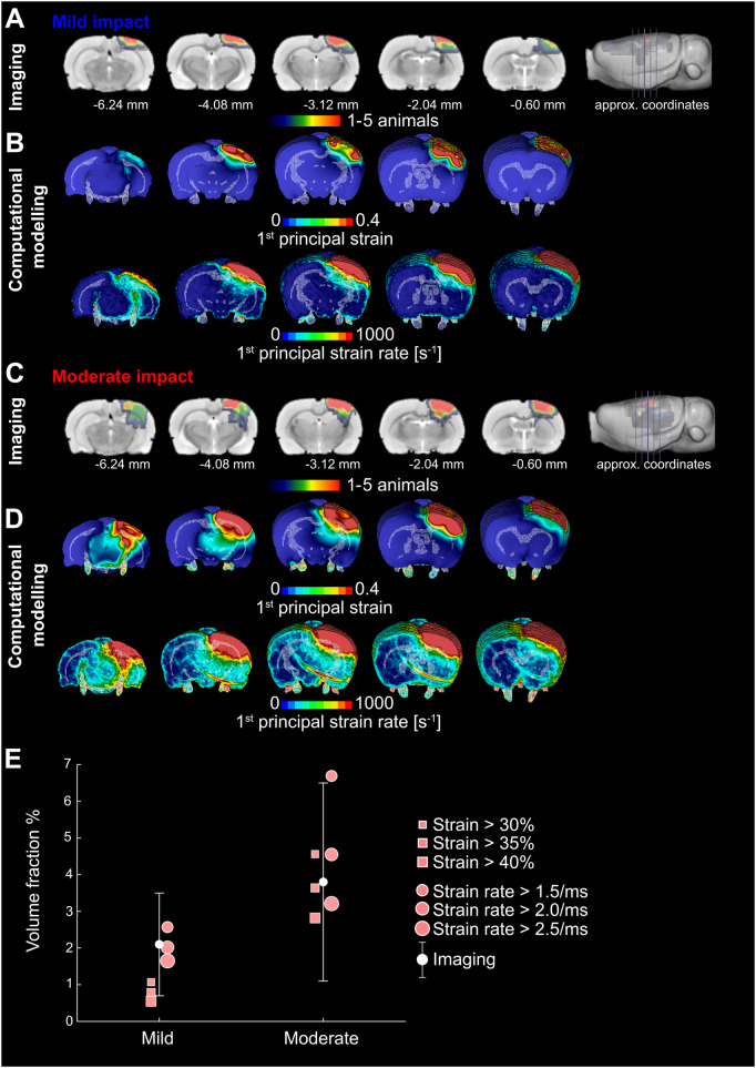
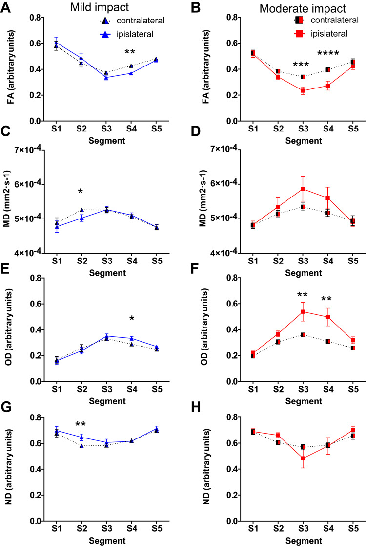
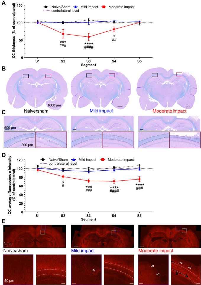
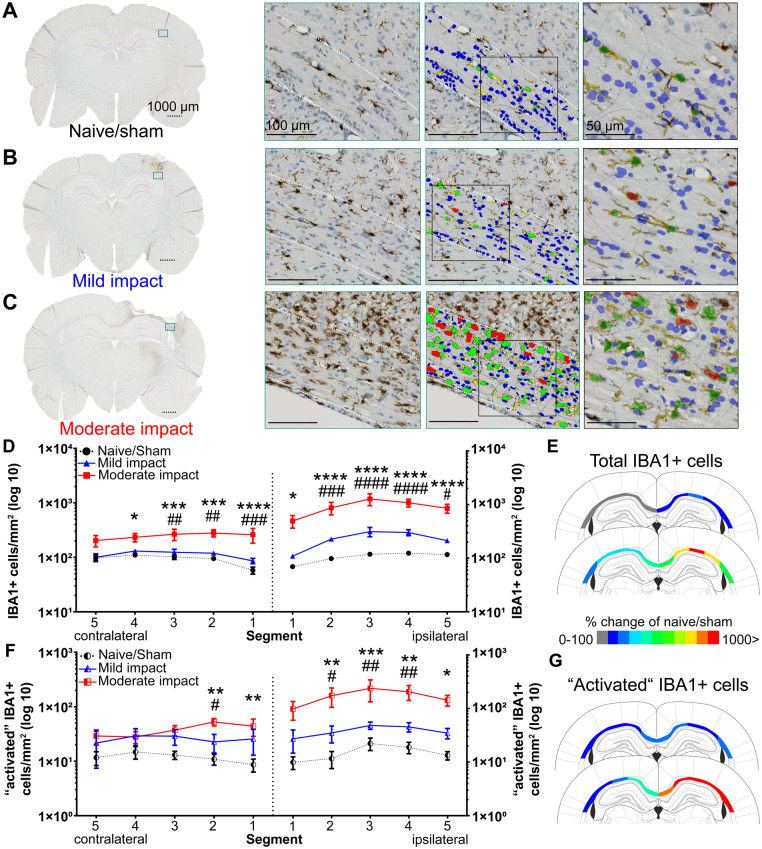
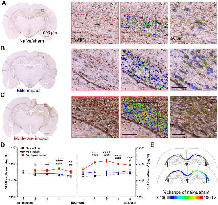
The relationship between biomechanical forces and neuropathology is key to understanding traumatic brain injury. White matter tracts are damaged by high shear forces during impact, resulting in axonal injury, a key determinant of long-term clinical outcomes. However, the relationship between biomechanical forces and patterns of white matter injuries, associated with persistent diffusion MRI abnormalities, is poorly understood. This limits the ability to predict the severity of head injuries and the design of appropriate protection. Our previously developed human finite element model of head injury predicted the location of post-traumatic neurodegeneration. A similar rat model now allows us to experimentally test whether strain patterns calculated by the model predicts in vivo MRI and histology changes. Using a controlled cortical impact, mild and moderate injuries (1 and 2 mm) were performed. Focal and axonal injuries were quantified with volumetric and diffusion 9.4 T MRI at 2 weeks post injury. Detailed analysis of the corpus callosum was conducted using multi-shell diffusion MRI and histopathology. Microglia and astrocyte density, including process parameters, along with white matter structural integrity and neurofilament expression were determined by quantitative immunohistochemistry. Linear mixed effects regression analyses for strain and strain rate with the employed outcome measures were used to ascertain how well immediate biomechanics could explain MRI and histology changes. The spatial pattern of mechanical strain and strain rate in the injured cortex shows good agreement with the probability maps of focal lesions derived from volumetric MRI. Diffusion metrics showed abnormalities in the corpus callosum, indicating white matter changes in the segments subjected to high strain, as predicted by the model. The same segments also exhibited a severity-dependent increase in glia cell density, white matter thinning and reduced neurofilament expression. Linear mixed effects regression analyses showed that mechanical strain and strain rate were significant predictors of in vivo MRI and histology changes. Specifically, strain and strain rate respectively explained 33% and 28% of the reduction in fractional anisotropy, 51% and 29% of the change in neurofilament expression and 51% and 30% of microglia density changes. The work provides evidence that strain and strain rate in the first milliseconds after injury are important factors in determining patterns of glial and axonal injury and serve as experimental validators of our computational model of traumatic brain injury. Our results provide support for the use of this model in understanding the relationship of biomechanics and neuropathology and can guide the development of head protection systems, such as airbags and helmets.
生物力学力与神经病理学之间的关系是理解创伤性脑损伤的关键。在冲击过程中,白质束会受到高剪切力的破坏,导致轴突损伤,这是长期临床结果的关键决定因素。然而,生物力学力与白质损伤模式之间的关系,与持续的扩散 MRI 异常有关,目前了解甚少。这限制了预测头部损伤严重程度和设计适当保护措施的能力。我们之前开发的头部损伤人类有限元模型预测了创伤后神经退行性变的位置。现在,类似的大鼠模型使我们能够通过实验测试模型计算的应变模式是否预测体内 MRI 和组织学变化。使用皮质控制冲击,进行轻度和中度损伤(1 和 2 毫米)。在损伤后 2 周使用容积和扩散 9.4 T MRI 定量检测局灶性和轴索性损伤。使用多壳扩散 MRI 和组织病理学对胼胝体进行详细分析。通过定量免疫组织化学确定小胶质细胞和星形胶质细胞密度,包括过程参数,以及白质结构完整性和神经丝表达。使用线性混合效应回归分析应变和应变速率与所采用的结果测量值之间的关系,以确定即时生物力学如何能够解释 MRI 和组织学变化。损伤皮质中的机械应变和应变速率的空间模式与源自容积 MRI 的局灶性病变概率图吻合良好。扩散指标显示胼胝体异常,表明模型预测的高应变段的白质变化。相同的节段还表现出神经胶质细胞密度、白质变薄和神经丝表达减少的严重程度依赖性增加。线性混合效应回归分析表明,机械应变和应变速率是体内 MRI 和组织学变化的重要预测因子。具体而言,应变和应变速率分别解释了各向异性分数降低的 33%和 28%,神经丝表达变化的 51%和 29%,以及小胶质细胞密度变化的 51%和 30%。这项工作提供了证据,证明损伤后最初几毫秒内的应变和应变速率是决定神经胶质和轴索性损伤模式的重要因素,并作为我们创伤性脑损伤计算模型的实验验证。我们的结果为使用该模型来理解生物力学和神经病理学之间的关系提供了支持,并可以指导头部保护系统(如安全气囊和头盔)的开发。