Fink Kathleen R, Fink James R
Department of Radiology, University of Washington, Seattle, WA 98104, USA.
Surg Neurol Int. 2013 May 2;4(Suppl 4):S209-19. doi: 10.4103/2152-7806.111298. Print 2013.
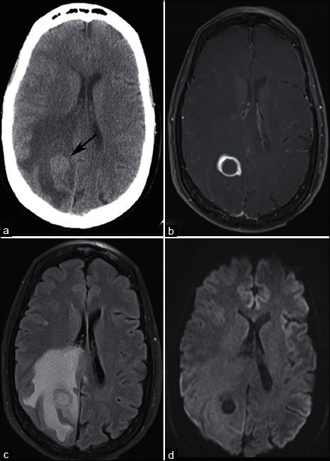
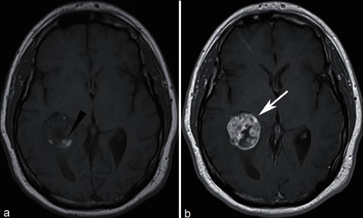
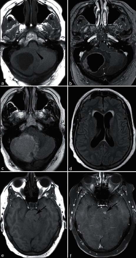
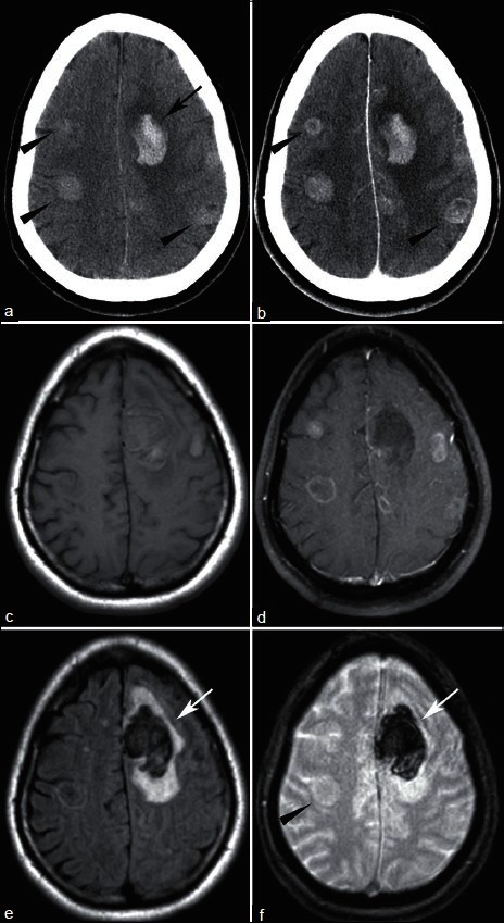
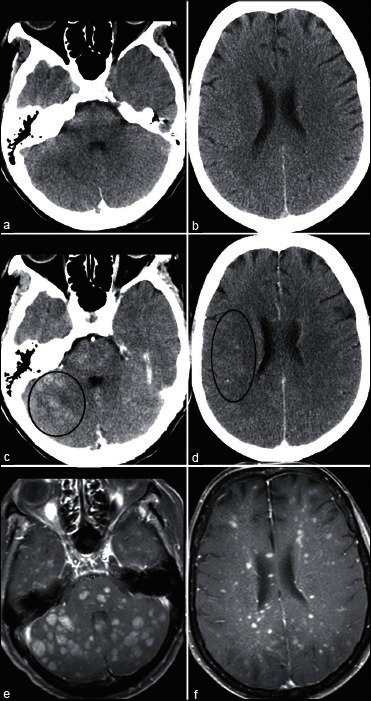
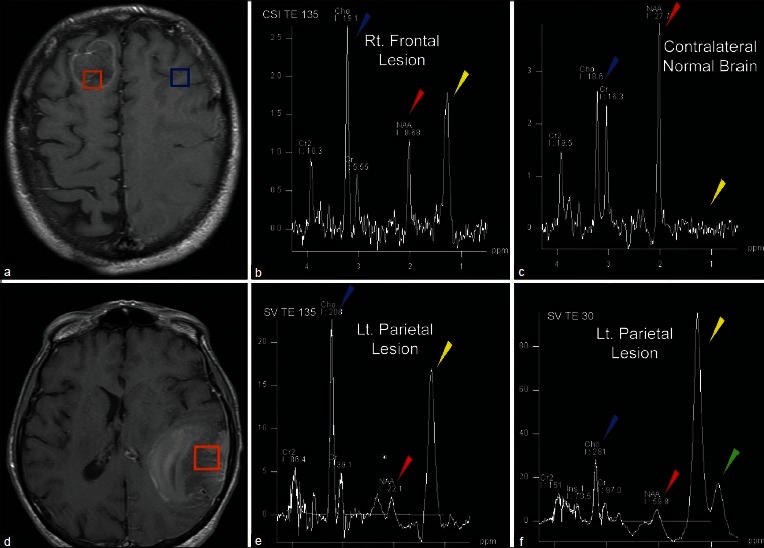
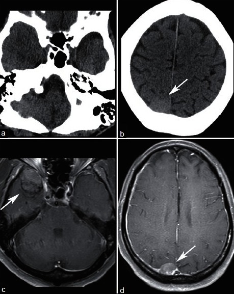
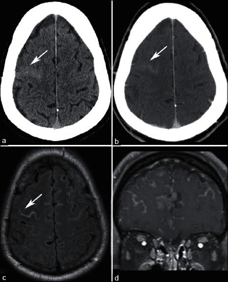
Imaging plays a key role in the diagnosis of central nervous system (CNS) metastasis. Imaging is used to detect metastases in patients with known malignancies and new neurological signs or symptoms, as well as to screen for CNS involvement in patients with known cancer. Computed tomography (CT) and magnetic resonance imaging (MRI) are the key imaging modalities used in the diagnosis of brain metastases. In difficult cases, such as newly diagnosed solitary enhancing brain lesions in patients without known malignancy, advanced imaging techniques including proton magnetic resonance spectroscopy (MRS), contrast enhanced magnetic resonance perfusion (MRP), diffusion weighted imaging (DWI), and diffusion tensor imaging (DTI) may aid in arriving at the correct diagnosis. This image-rich review discusses the imaging evaluation of patients with suspected intracranial involvement and malignancy, describes typical imaging findings of parenchymal brain metastasis on CT and MRI, and provides clues to specific histological diagnoses such as the presence of hemorrhage. Additionally, the role of advanced imaging techniques is reviewed, specifically in the context of differentiating metastasis from high-grade glioma and other solitary enhancing brain lesions. Extra-axial CNS involvement by metastases, including pachymeningeal and leptomeningeal metastases is also briefly reviewed.
影像学在中枢神经系统(CNS)转移瘤的诊断中起着关键作用。影像学用于检测已知患有恶性肿瘤且出现新的神经体征或症状的患者的转移瘤,以及筛查已知患有癌症的患者的中枢神经系统受累情况。计算机断层扫描(CT)和磁共振成像(MRI)是用于诊断脑转移瘤的关键影像学检查方法。在一些疑难病例中,例如在没有已知恶性肿瘤的患者中新诊断出的孤立性强化脑病变,包括质子磁共振波谱(MRS)、对比增强磁共振灌注(MRP)、扩散加权成像(DWI)和扩散张量成像(DTI)等先进的影像学技术可能有助于做出正确诊断。这篇内容丰富的综述讨论了疑似颅内受累和恶性肿瘤患者的影像学评估,描述了脑实质转移瘤在CT和MRI上的典型影像学表现,并提供了诸如出血存在等特定组织学诊断的线索。此外,还综述了先进影像学技术的作用,特别是在区分转移瘤与高级别胶质瘤及其他孤立性强化脑病变方面。还简要回顾了转移瘤对轴外中枢神经系统的累及情况,包括硬脑膜和软脑膜转移。