Department of Operative Dentistry, University of Pavia, Policlinico "San Matteo", Piazzale Golgi 3, 27100 Pavia, Italy,
Med Oral Patol Oral Cir Bucal. 2013 Sep 1;18(5):e793-8. doi: 10.4317/medoral.18344.
The purpose of this in vitro study was to evaluate the microleakage in "deep" Class II composite restorations with gingival cavosurface margin below the CEJ (cemento-enamel junction) and restored with different techniques.
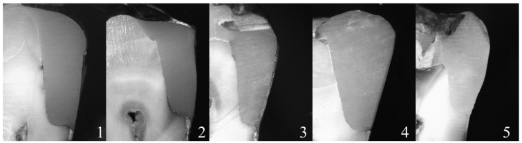
Fifty human teeth were used. In each tooth two standardized Class II slot cavities (on mesial and on distal surfaces) were prepared: the buccolingual extension of the cavities was 4 mm; the gingival wall was located in dentin/cementum (2 mm beyond the CEJ). The prepared teeth were randomly assigned to 5 experimental groups (of 10 specimens and 20 cavities each) and restored. Group 1: Filtek TM Supreme XTE Flowable (3MESPE) + Universal Filtek Supreme XTE (3MESPE), Group 2: GrandioSO Heavy Flow (Voco) + GrandioSo (Voco), Group 3: SDR™ (Dentsply Caulk) + Esthet-X® HD (Dentsply Caulk), Group 4: SonicFill (Kerr), Group 5: Grandio (Voco). After thermocycling, the specimens were immersed in a 0.5% basic fuchsine dye solution and incubated at 37°C for 24 hours. The teeth were subsequently sectioned mesiodistally. All specimens were examined at 25 in a stereomicroscope and standardized digital images were obtained. Dye penetration was measured from gingival margins.
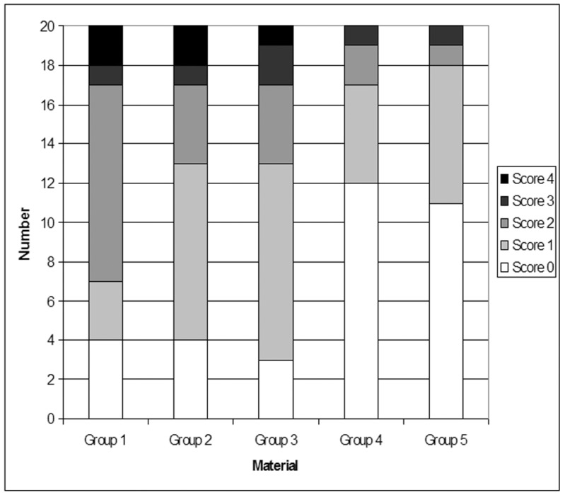
The results demonstrated no significant leakage differences between Group 4 and Group 5, that both showed significantly higher frequency distribution of Score 0. Group 2 and Group 3 showed a significant prevalence of Score 1, whereas Group 1 showed significantly higher frequency of Score 2.
None of the restorative techniques tested completely eliminated microleakage dye penetration in dentin margins; marginal adaptation in Class II composite restorations with gingival wall below the CEJ varied in both substrates and from different restorative techniques used.
本体外研究的目的是评估在 CEJ(牙釉质-牙骨质界)以下龈壁的“深”Ⅱ类复合修复中不同技术的微渗漏。
本研究共使用了 50 个人工牙。每个牙制备两个标准化的Ⅱ类槽状窝(在近中和远中面):窝洞的颊舌向扩展为 4mm;龈壁位于牙本质/牙骨质(在 CEJ 后 2mm)。制备后的牙齿随机分为 5 个实验组(每组 10 个标本,20 个窝洞)并进行修复。第 1 组:Filtek TM Supreme XTE Flowable(3MESPE)+Universal Filtek Supreme XTE(3MESPE),第 2 组:GrandioSO Heavy Flow(Voco)+GrandioSo(Voco),第 3 组:SDR™(Dentsply Caulk)+Esthet-X® HD(Dentsply Caulk),第 4 组:SonicFill(Kerr),第 5 组:Grandio(Voco)。热循环后,将标本浸泡在 0.5%碱性碱性品红染料溶液中,在 37°C 下孵育 24 小时。随后将牙齿沿近远中方向切割。所有标本在立体显微镜下以 25°角检查,并获得标准化的数字图像。从龈缘测量染料渗透。
结果显示,第 4 组和第 5 组之间的微渗漏差异无统计学意义,两组均显示出明显更高的 Score 0 分布频率。第 2 组和第 3 组的 Score 1 发生率显著较高,而第 1 组的 Score 2 发生率显著较高。
在所测试的修复技术中,没有一种技术能完全消除牙本质边缘的复合修复微渗漏染料渗透;在 CEJ 以下龈壁的Ⅱ类复合修复中,边缘适应性在不同的基底和使用的不同修复技术中都存在差异。