Department of Diagnostic Imaging and Interventional Radiology, Pomeranian Medical University, Clinical Hospital No. 1, Unii Lubelskiej 1, Szczecin, 71252, Poland.
Neuroradiology. 2013 Sep;55(9):1061-9. doi: 10.1007/s00234-013-1210-5. Epub 2013 Jun 2.
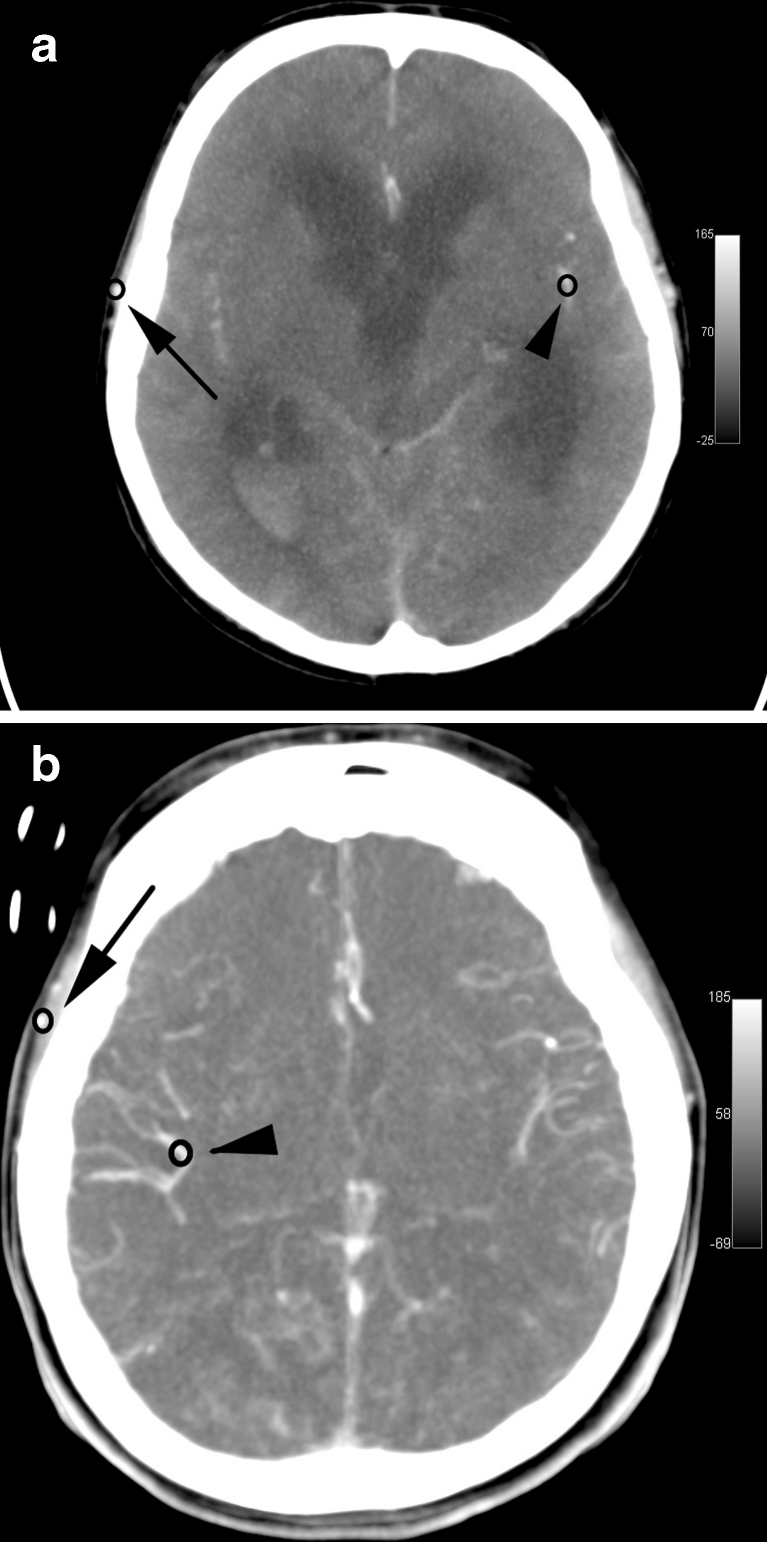
Stasis filling, defined as delayed, weak, and persistent opacification of proximal segments of the cerebral arteries, is frequently found in brain dead patients. This phenomenon causes a major problem in the development of reliable computed tomographic angiography (CTA) protocol in the diagnosis of brain death (BD). The aim of our study was to characterize stasis filling in the diagnosis of BD. To achieve this, we performed a dynamic evaluation of contrast enhancement of the cerebral and extracranial arteries in patients with BD and controls.
Study population included 30 BD patients, who showed stasis filling in computed tomographic perfusion (CTP) series. Thirty patients, after clipping of an intracranial aneurysm, constituted the control group. The study protocol consisted of CTA, CTP, and angiography. Time-density curves (TDCs) of cerebral and extracranial arteries were generated using 40-s series of CTP.
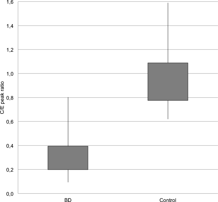
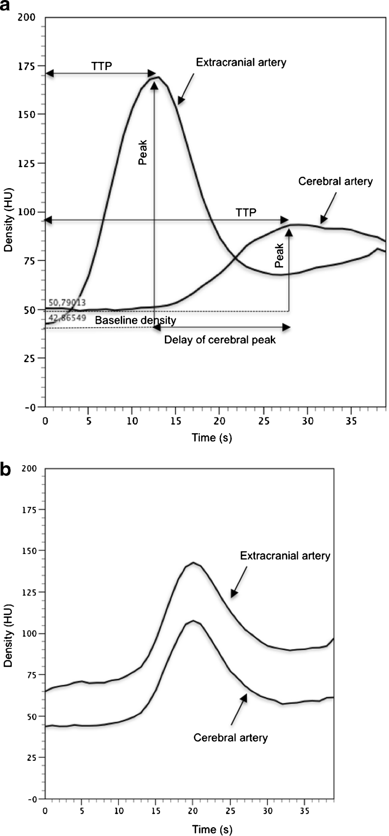
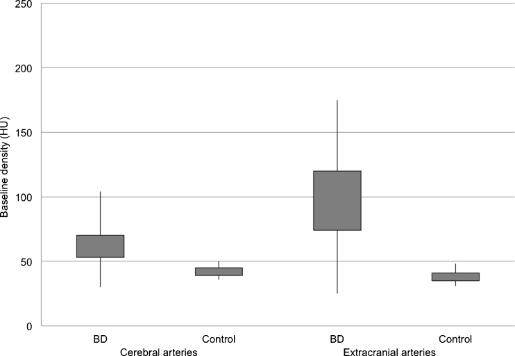
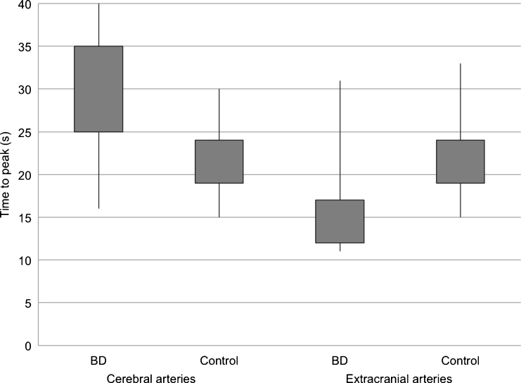
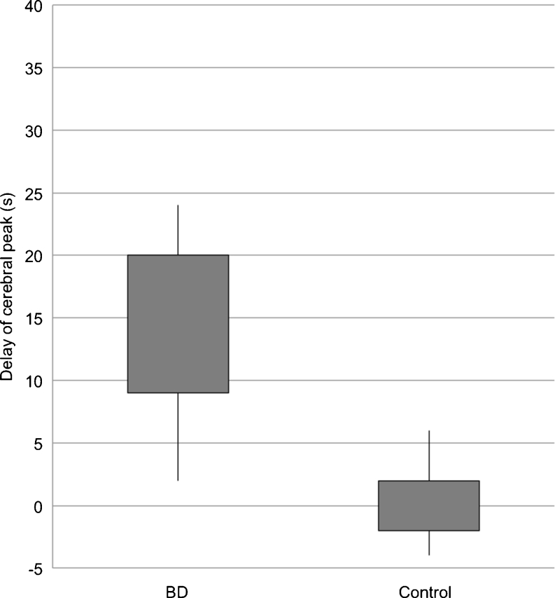
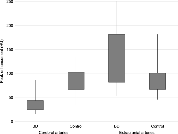
Cerebral TDCs in BD patients represented flat curves in contrast to TDCs in controls, which formed steep and narrow Gaussian curves. We found longer time to peak enhancement in BD patients than in controls (32 vs. 21 s; p < 0.0001). In BD patients, peak enhancement in the cerebral arteries occurred with a median delay of 14.5 s to peak in extracranial arteries, while no delay was noted in controls (p < 0.0001). Cerebral arteries in BD patients showed lower peak enhancement than controls (34.5 vs. 81.5 HU; p < 0.0001). In all BD patients, CTP revealed zero values of cerebral blood flow and volume. Angiography showed stasis filling in 14 (46.7 %) and non-filling in 16 (53.3 %) cases.
A confrontation of stasis filling with CTP results showed that stasis filling is not consistent with preserved cerebral perfusion, thus does not preclude diagnosis of BD.
在脑死亡患者中,经常可以发现血流停滞现象,即近端脑动脉的延迟、微弱和持续显影。这种现象在脑死亡(BD)的计算机断层血管造影(CTA)诊断中造成了一个主要问题。我们的研究旨在对脑死亡诊断中的血流停滞现象进行特征描述。为此,我们对脑死亡患者和对照组患者进行了脑和颅外动脉对比增强的动态评估。
研究人群包括 30 名在 CT 灌注(CTP)系列中显示血流停滞的脑死亡患者。另外 30 名接受颅内动脉瘤夹闭的患者构成对照组。研究方案包括 CTA、CTP 和血管造影。通过 CTP 的 40 秒系列生成脑和颅外动脉的时间密度曲线(TDC)。
与对照组相比,脑死亡患者的脑 TDC 呈平坦曲线,而对照组的 TDC 呈陡峭而狭窄的高斯曲线。我们发现脑死亡患者的峰值增强时间比对照组长(32 对 21 秒;p < 0.0001)。在脑死亡患者中,脑动脉的峰值增强发生在外侧动脉峰值增强后中位数延迟 14.5 秒,而对照组没有延迟(p < 0.0001)。脑死亡患者的脑动脉峰值增强低于对照组(34.5 对 81.5 HU;p < 0.0001)。在所有脑死亡患者中,CTP 显示脑血流和容积均为零值。血管造影显示 14 例(46.7%)存在血流停滞,16 例(53.3%)不存在血流停滞。
将血流停滞与 CTP 结果进行对比发现,血流停滞与脑灌注的保留不一致,因此不能排除脑死亡的诊断。