Focal Area Infection Biology, Biozentrum, University of Basel, Basel, Switzerland.
PLoS One. 2013 May 30;8(5):e64901. doi: 10.1371/journal.pone.0064901. Print 2013.
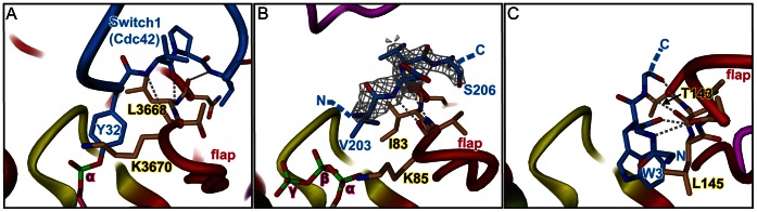
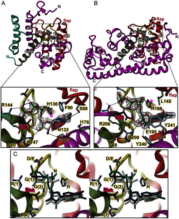
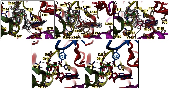
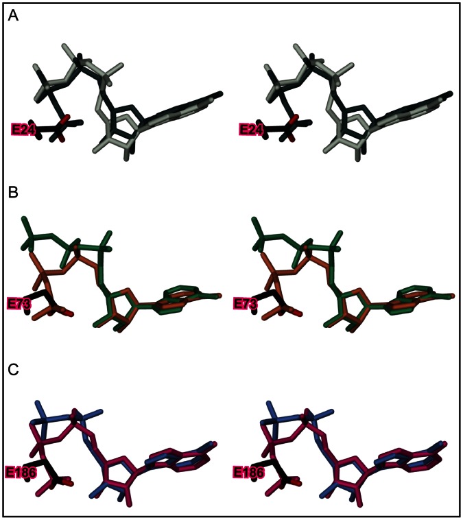
The ubiquitous FIC domain is evolutionarily conserved from bacteria to human and has been shown to catalyze AMP transfer onto protein side-chain hydroxyl groups. Recently, it was predicted that most catalytically competent Fic proteins are inhibited by the presence of an inhibitory helix αinh that is provided by a cognate anti-toxin (class I), or is part of the N- or C-terminal part of the Fic protein itself (classes II and III). In vitro, inhibition is relieved by mutation of a conserved glutamate of αinh to glycine. For the class III bacterial Fic protein NmFic from Neisseria meningitidis, the inhibitory mechanism has been elucidated. Here, we extend above study by including bacterial class I and II Fic proteins VbhT from Bartonella schoenbuchensis and SoFic from Shewanella oneidensis, respectively, and the respective E->G mutants. Comparative enzymatic and crystallographic analyses show that, in all three classes, the ATP substrate binds to the wild-type FIC domains, but with the α-phosphate in disparate and non-competent orientations. In the E->G mutants, however, the tri-phosphate moiety is found reorganized to the same tightly bound structure through a unique set of hydrogen bonds with Fic signature motif residues. The γ-phosphate adopts the location that is taken by the inhibitory glutamate in wild-type resulting in an α-phosphate orientation that can be attacked in-line by a target side-chain hydroxyl group. The latter is properly registered to the Fic active center by main-chain β-interactions with the β-hairpin flap. These data indicate that the active site motif and the exposed edge of the flap are both required to form an adenylylation-competent Fic protein.
普遍存在的 FIC 结构域在从细菌到人等生物中都具有进化保守性,并且已被证明可催化 AMP 转移到蛋白质侧链羟基上。最近,有人预测,大多数具有催化能力的 Fic 蛋白都受到抑制性αinh 螺旋的抑制,该螺旋由同源的抗毒素(I 类)提供,或者是 Fic 蛋白自身的 N 端或 C 端的一部分(II 类和 III 类)。在体外,通过突变αinh 中保守的谷氨酸为甘氨酸可以解除抑制。对于脑膜炎奈瑟菌的 III 类细菌 Fic 蛋白 NmFic,已阐明了其抑制机制。在这里,我们通过包括来自巴尔通体属的 Bartonella schoenbuchensis 的 VbhT 和来自希瓦氏菌属的 Shewanella oneidensis 的 SoFic 以及各自的 E->G 突变体,扩展了上述研究。比较酶学和晶体学分析表明,在所有三类 FIC 结构域中,ATP 底物都与野生型 FIC 结构域结合,但α-磷酸的位置不同且不具有竞争能力。然而,在 E->G 突变体中,三磷酸部分通过与 Fic 特征基序残基形成独特的氢键重新组织成相同的紧密结合结构。γ-磷酸占据了野生型中抑制性谷氨酸的位置,从而使α-磷酸的取向可以通过靶标侧链羟基的直线进攻。后者通过与β发夹瓣的主链β-相互作用,正确地注册到 Fic 活性中心,从而实现腺苷酰化的活性。这些数据表明,活性位点基序和瓣的暴露边缘都需要形成具有腺苷酰化能力的 Fic 蛋白。