• 文献检索
  • 文档翻译
  • 深度研究
  • 学术资讯
  • Suppr Zotero 插件Zotero 插件
  • 邀请有礼
  • 套餐&价格
  • 历史记录
应用&插件
Suppr Zotero 插件Zotero 插件浏览器插件Mac 客户端Windows 客户端微信小程序
定价
高级版会员购买积分包购买API积分包
服务
文献检索文档翻译深度研究API 文档MCP 服务
关于我们
关于 Suppr公司介绍联系我们用户协议隐私条款
关注我们

Suppr 超能文献

核心技术专利:CN118964589B侵权必究
粤ICP备2023148730 号-1Suppr @ 2026

文献检索

告别复杂PubMed语法,用中文像聊天一样搜索,搜遍4000万医学文献。AI智能推荐,让科研检索更轻松。

立即免费搜索

文件翻译

保留排版,准确专业,支持PDF/Word/PPT等文件格式,支持 12+语言互译。

免费翻译文档

深度研究

AI帮你快速写综述,25分钟生成高质量综述,智能提取关键信息,辅助科研写作。

立即免费体验

椎动脉损伤中颈椎骨折脱位的机制与模式

Mechanism and patterns of cervical spine fractures-dislocations in vertebral artery injury.

作者信息

Gupta Pankaj, Kumar Atin, Gamangatti Shivanand

机构信息

Department of Radiodiagnosis, All India Institute of Medical Sciences, New Delhi, India.

出版信息

J Craniovertebr Junction Spine. 2012 Jan;3(1):11-5. doi: 10.4103/0974-8237.110118.

DOI:10.4103/0974-8237.110118
PMID:23741123
原文链接:https://pmc.ncbi.nlm.nih.gov/articles/PMC3669467/
Abstract

PURPOSE

To identify the fracture patterns and mechanism of injury, based on subaxial cervical spine injury classification system (SLIC), on non-contrast computed tomography (NCCT) of cervical spine predictive of vertebral artery injury (VAI).

PATIENTS AND METHODS

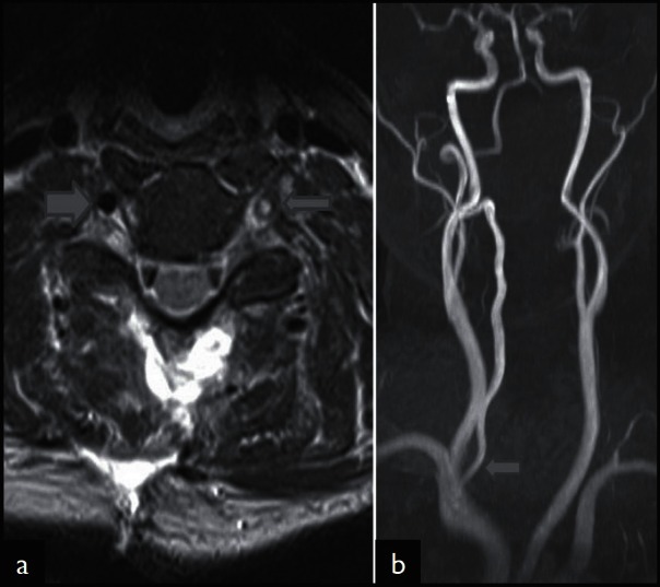
We retrospectively analyzed cervical spine magnetic resonance imaging (MRI) of 320 patients who were admitted with cervical spine injury in our level I regional trauma center over a period of two years (April 2010 to April 2012). Diagnosis of VAI was based on hyperintensity replacing the flow void on a T2-weighted axial image. NCCT images of the selected 43 patients with MRI diagnosis of VAI were then assessed for the pattern of injury. The cervical spinal injuries were classified into those involving the C1 and C2 and subaxial spine. For the latter, SLIC was used.

RESULTS

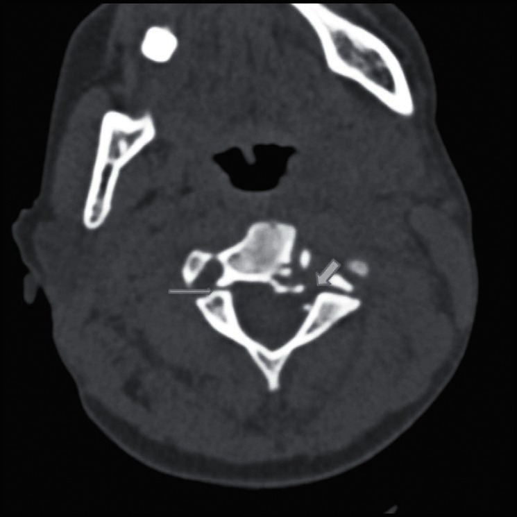
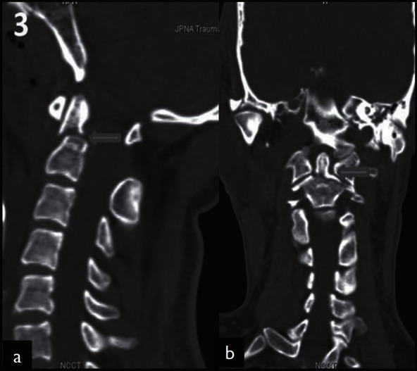
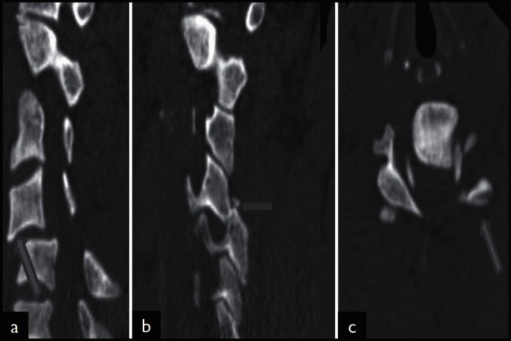
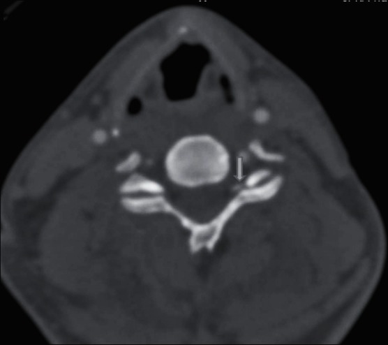
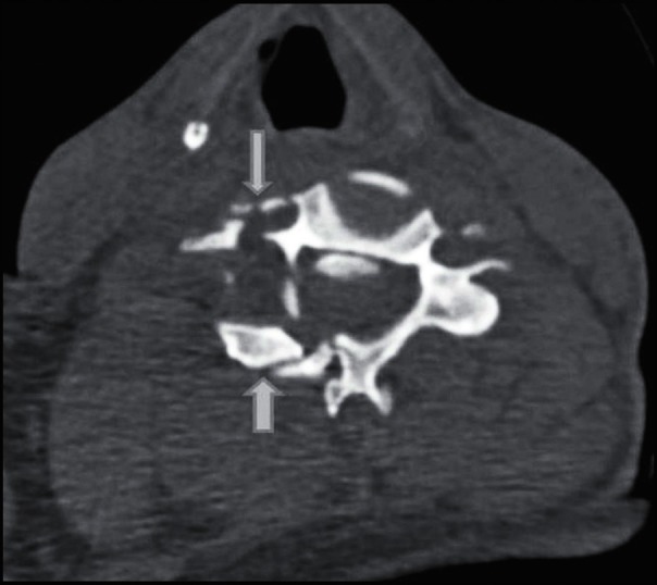
A total of 47 VAI were analyzed in 43 patients. Only one patient with VAI on MRI had no detectable abnormality on NCCT. C1 and C2 injuries were found in one and six patients respectively. In subaxial injuries, the most common mechanism of injury was distraction (37.5%) with facet dislocation with or without fracture representing the most common pattern of injury (55%). C5 was the single most common affected vertebral level. Extension to foramen transversarium was present in 20 (42.5%) cases.

CONCLUSION

CT represents a robust screening tool for patients with VAI. VAI should be suspected in patients with facet dislocation with or without fractures, foramina transversarium fractures and C1-C3 fractures, especially type III odontoid fractures and distraction mechanism of injury.

摘要

目的

基于下颈椎损伤分类系统(SLIC),在颈椎非增强计算机断层扫描(NCCT)上确定骨折类型及损伤机制,以预测椎动脉损伤(VAI)。

患者与方法

我们回顾性分析了我院一级区域创伤中心在两年时间(2010年4月至2012年4月)内收治的320例颈椎损伤患者的颈椎磁共振成像(MRI)。VAI的诊断基于T2加权轴位图像上血流空洞被高信号取代。然后对43例经MRI诊断为VAI的患者的NCCT图像进行损伤类型评估。颈椎损伤分为涉及C1和C2的损伤以及下颈椎损伤。对于后者,采用SLIC分类。

结果

共分析了43例患者的47处VAI。MRI显示VAI的患者中,只有1例在NCCT上未发现可检测到的异常。分别有1例和6例患者存在C1和C2损伤。在下颈椎损伤中,最常见的损伤机制是牵张(37.5%),伴或不伴骨折的小关节脱位是最常见的损伤类型(55%)。C5是最常受累的单个椎体节段。20例(42.5%)病例存在横突孔延伸损伤。

结论

CT是VAI患者的一种可靠筛查工具。对于伴或不伴骨折的小关节脱位、横突孔骨折和C1 - C3骨折患者,尤其是III型齿状突骨折和牵张损伤机制的患者,应怀疑存在VAI。

https://cdn.ncbi.nlm.nih.gov/pmc/blobs/38a8/3669467/d859c18b326c/JCVJS-3-11-g006.jpg
https://cdn.ncbi.nlm.nih.gov/pmc/blobs/38a8/3669467/cf59b3c0dde2/JCVJS-3-11-g001.jpg
https://cdn.ncbi.nlm.nih.gov/pmc/blobs/38a8/3669467/0e57f7557927/JCVJS-3-11-g002.jpg
https://cdn.ncbi.nlm.nih.gov/pmc/blobs/38a8/3669467/3a7a130625e7/JCVJS-3-11-g003.jpg
https://cdn.ncbi.nlm.nih.gov/pmc/blobs/38a8/3669467/d180f22d6579/JCVJS-3-11-g004.jpg
https://cdn.ncbi.nlm.nih.gov/pmc/blobs/38a8/3669467/cc8b9a8888f7/JCVJS-3-11-g005.jpg
https://cdn.ncbi.nlm.nih.gov/pmc/blobs/38a8/3669467/d859c18b326c/JCVJS-3-11-g006.jpg

相似文献

1
Mechanism and patterns of cervical spine fractures-dislocations in vertebral artery injury.椎动脉损伤中颈椎骨折脱位的机制与模式
J Craniovertebr Junction Spine. 2012 Jan;3(1):11-5. doi: 10.4103/0974-8237.110118.
2
Vertebral Artery Injury in Cervical Spine Fracture Dislocation: An Observational Study in a Tertiary Care Center.颈椎骨折脱位中的椎动脉损伤:在一家三级医疗中心的观察性研究
Cureus. 2024 Apr 7;16(4):e57774. doi: 10.7759/cureus.57774. eCollection 2024 Apr.
3
Cervical spine fracture patterns predictive of blunt vertebral artery injury.预测钝性椎动脉损伤的颈椎骨折类型
J Trauma. 2003 Nov;55(5):811-3. doi: 10.1097/01.TA.0000092700.92587.32.
4
Cervical spine fracture patterns mandating screening to rule out blunt cerebrovascular injury.需要进行筛查以排除钝性脑血管损伤的颈椎骨折类型。
Surgery. 2007 Jan;141(1):76-82. doi: 10.1016/j.surg.2006.04.005. Epub 2006 Aug 28.
5
Correlation of C2 fractures and vertebral artery injury.C2 骨折与椎动脉损伤的相关性。
Spine (Phila Pa 1976). 2010 May 20;35(12):E520-4. doi: 10.1097/BRS.0b013e3181cd98b6.
6
Incidence and Outcome Analysis of Vertebral Artery Injury in Posttraumatic Cervical Spine.创伤性颈椎椎动脉损伤的发病率及转归分析
Asian J Neurosurg. 2020 Aug 28;15(3):644-647. doi: 10.4103/ajns.AJNS_45_20. eCollection 2020 Jul-Sep.
7
Predictors of Vertebral Artery Injury in Isolated C2 Fractures Based on Fracture Morphology Using CT Angiography.基于CT血管造影术骨折形态学的孤立性C2骨折椎动脉损伤预测因素
Spine (Phila Pa 1976). 2015 Jun 15;40(12):E713-8. doi: 10.1097/BRS.0000000000000893.
8
Vertebral artery injury associated with blunt cervical spine trauma: a multivariate regression analysis.与钝性颈椎创伤相关的椎动脉损伤:多变量回归分析。
Spine (Phila Pa 1976). 2013 Jul 15;38(16):1352-61. doi: 10.1097/BRS.0b013e318294bacb.
9
Vertebral artery injury in destabilized midcervical spine trauma; predisposing factors and proposed mechanism.不稳定型颈中脊柱创伤中的椎动脉损伤;易患因素和拟议的发病机制。
Acta Neurochir (Wien). 2012 Nov;154(11):2091-8; discussion 2098. doi: 10.1007/s00701-012-1499-6. Epub 2012 Sep 19.
10
Screening via CT angiogram after traumatic cervical spine fractures: narrowing imaging to improve cost effectiveness. Experience of a Level I trauma center.创伤性颈椎骨折后通过CT血管造影进行筛查:缩小成像范围以提高成本效益。一级创伤中心的经验。
J Neurosurg Spine. 2016 Mar;24(3):490-5. doi: 10.3171/2015.6.SPINE15140. Epub 2015 Nov 27.

引用本文的文献

1
Traumatic Vertebral Artery Injury: Diagnosis, Natural History, and Key Considerations for Management.创伤性椎动脉损伤:诊断、自然病程及管理的关键考量因素
J Clin Med. 2025 May 2;14(9):3159. doi: 10.3390/jcm14093159.
2
Clinical characteristics and outcomes of patients with concomitant cervical spine trauma and vertebral artery injury: A literature review and retrospective analysis.颈椎创伤合并椎动脉损伤患者的临床特征与预后:文献综述及回顾性分析
J Craniovertebr Junction Spine. 2025 Jan-Mar;16(1):47-53. doi: 10.4103/jcvjs.jcvjs_207_24. Epub 2025 Apr 1.
3
Don't Hang Around, It Could Be Incidental: A Case Report of Hangman's Fracture and Review of the Literature.

本文引用的文献

1
Vertebral artery injury after cervical spine trauma: A prospective study using computed tomographic angiography.颈椎创伤后椎动脉损伤:一项使用计算机断层血管造影的前瞻性研究。
Surg Neurol Int. 2011 Mar 23;2:39. doi: 10.4103/2152-7806.78255.
2
Blunt cerebrovascular injury practice management guidelines: the Eastern Association for the Surgery of Trauma.钝性脑血管损伤实践管理指南:东部创伤外科学会
J Trauma. 2010 Feb;68(2):471-7. doi: 10.1097/TA.0b013e3181cb43da.
3
Does the morphology of foramen transversarium fractures predict vertebral artery injuries?
别磨蹭了,这可能是偶然发现的:一例绞刑者骨折病例报告及文献综述
Cureus. 2024 Jun 27;16(6):e63285. doi: 10.7759/cureus.63285. eCollection 2024 Jun.
4
Vertebral Artery Injury in Cervical Spine Fracture Dislocation: An Observational Study in a Tertiary Care Center.颈椎骨折脱位中的椎动脉损伤:在一家三级医疗中心的观察性研究
Cureus. 2024 Apr 7;16(4):e57774. doi: 10.7759/cureus.57774. eCollection 2024 Apr.
5
The value of magnetic resonance imaging and computed tomography in the study of spinal disorders.磁共振成像和计算机断层扫描在脊柱疾病研究中的价值。
Quant Imaging Med Surg. 2022 Jul;12(7):3947-3986. doi: 10.21037/qims-2022-04.
6
Minimally displaced unilateral facet fracture of cervical spine can lead to spinal cord injury: a report of two cases.颈椎轻度移位的单侧小关节骨折可导致脊髓损伤:两例报告。
BMC Musculoskelet Disord. 2021 Feb 11;22(1):168. doi: 10.1186/s12891-021-04025-x.
7
Incidence and Outcome Analysis of Vertebral Artery Injury in Posttraumatic Cervical Spine.创伤性颈椎椎动脉损伤的发病率及转归分析
Asian J Neurosurg. 2020 Aug 28;15(3):644-647. doi: 10.4103/ajns.AJNS_45_20. eCollection 2020 Jul-Sep.
横突孔骨折的形态学是否能预测椎动脉损伤?
Spine (Phila Pa 1976). 2008 Dec 1;33(25):E957-61. doi: 10.1097/BRS.0b013e31818e2f31.
4
ACR Appropriateness Criteria on suspected spine trauma.美国放射学会关于疑似脊柱创伤的适宜性标准
J Am Coll Radiol. 2007 Nov;4(11):762-75. doi: 10.1016/j.jacr.2007.08.006.
5
The subaxial cervical spine injury classification system: a novel approach to recognize the importance of morphology, neurology, and integrity of the disco-ligamentous complex.下颈椎损伤分类系统:一种认识椎间盘韧带复合体形态、神经学及完整性重要性的新方法。
Spine (Phila Pa 1976). 2007 Oct 1;32(21):2365-74. doi: 10.1097/BRS.0b013e3181557b92.
6
Cervical spine fracture patterns mandating screening to rule out blunt cerebrovascular injury.需要进行筛查以排除钝性脑血管损伤的颈椎骨折类型。
Surgery. 2007 Jan;141(1):76-82. doi: 10.1016/j.surg.2006.04.005. Epub 2006 Aug 28.
7
Sixteen-slice multi-detector computed tomographic angiography improves the accuracy of screening for blunt cerebrovascular injury.16层多探测器计算机断层血管造影术提高了钝性脑血管损伤筛查的准确性。
J Trauma. 2006 Jun;60(6):1204-9; discussion 1209-10. doi: 10.1097/01.ta.0000220435.55791.ce.
8
Helical computed tomographic angiography: an excellent screening test for blunt cerebrovascular injury.螺旋计算机断层血管造影术:钝性脑血管损伤的极佳筛查方法。
J Trauma. 2004 Jul;57(1):11-7; discussion 17-9. doi: 10.1097/01.ta.0000135499.70048.c7.
9
Vertebral artery in relationship to C1-C2 vertebrae: an anatomical study.椎动脉与C1-C2椎体的关系:一项解剖学研究。
Neurol India. 2004 Jun;52(2):178-84.
10
Cervical spine fracture patterns predictive of blunt vertebral artery injury.预测钝性椎动脉损伤的颈椎骨折类型
J Trauma. 2003 Nov;55(5):811-3. doi: 10.1097/01.TA.0000092700.92587.32.