Research Division, Joslin Diabetes Center and Harvard Medical School, Boston, Massachusetts, USA.
Diabetes. 2013 Sep;62(9):3081-92. doi: 10.2337/db12-1261. Epub 2013 Jun 12.
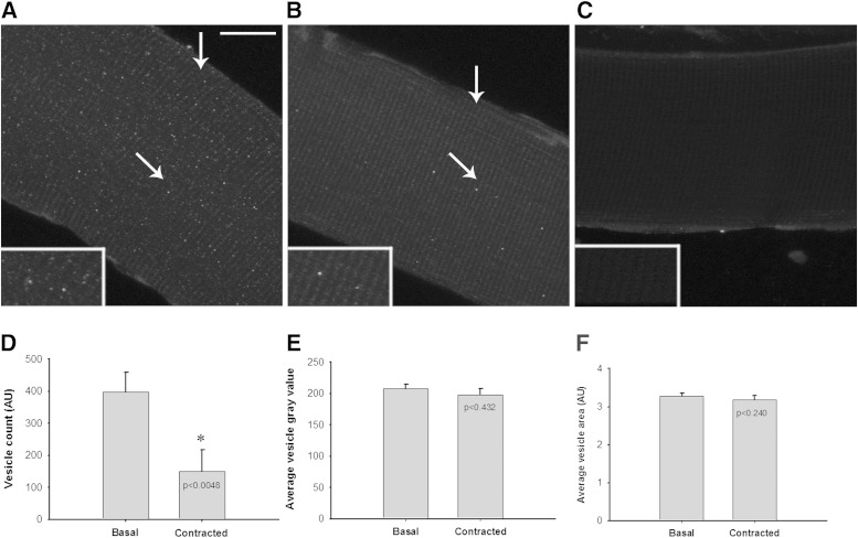
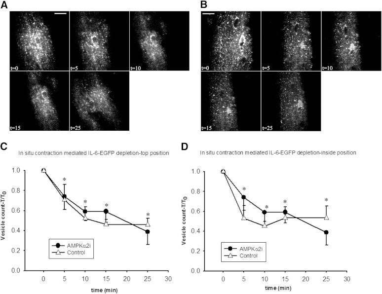
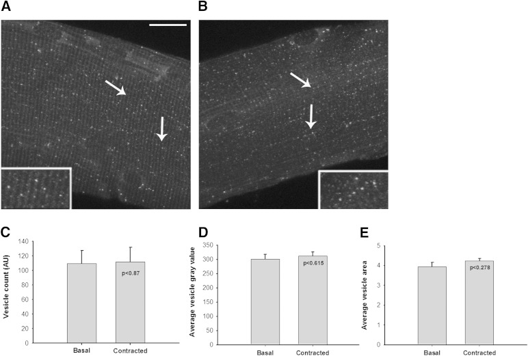
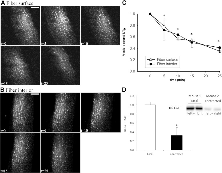
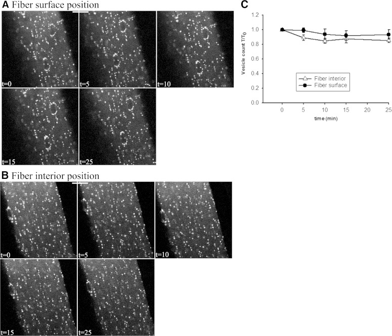
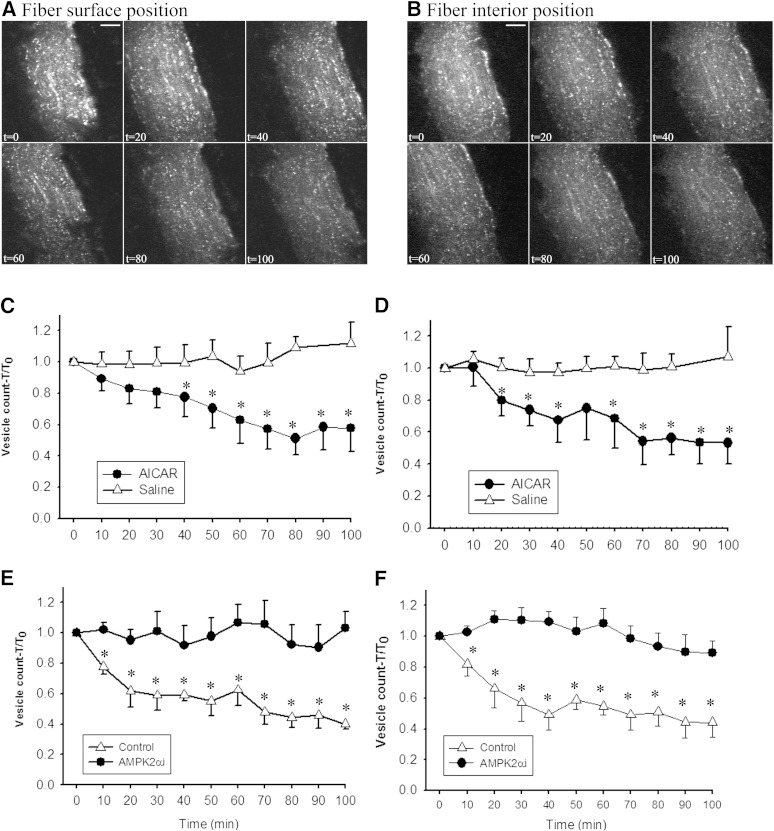
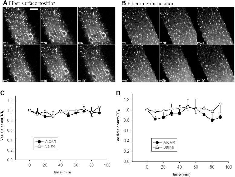
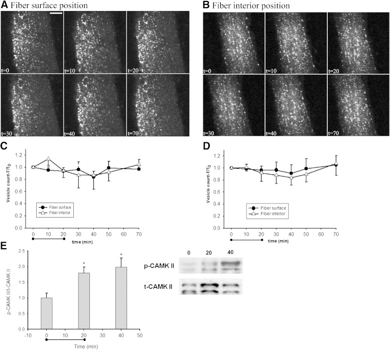
Recent studies suggest that interleukin 6 (IL-6) is released from contracting skeletal muscles; however, the cellular origin, secretion kinetics, and signaling mechanisms regulating IL-6 secretion are unknown. To address these questions, we developed imaging methodology to study IL-6 in fixed mouse muscle fibers and in live animals in vivo. Using confocal imaging to visualize endogenous IL-6 protein in fixed muscle fibers, we found IL-6 in small vesicle structures distributed throughout the fibers under basal (resting) conditions. To determine the kinetics of IL-6 secretion, intact quadriceps muscles were transfected with enhanced green fluorescent protein (EGFP)-tagged IL-6 (IL-6-EGFP), and 5 days later anesthetized mice were imaged before and after muscle contractions in situ. Contractions decreased IL-6-EGFP-containing vesicles and protein by 62% (P < 0.05), occurring rapidly and progressively over 25 min of contraction. However, contraction-mediated IL-6-EGFP reduction was normal in muscle-specific AMP-activated protein kinase (AMPK) α2-inactive transgenic mice. In contrast, the AMPK activator AICAR decreased IL-6-EGFP vesicles, an effect that was inhibited in the transgenic mice. In conclusion, resting skeletal muscles contain IL-6-positive vesicles that are expressed throughout myofibers. Contractions stimulate the rapid reduction of IL-6 in myofibers, occurring through an AMPKα2-independent mechanism. This novel imaging methodology clearly establishes IL-6 as a contraction-stimulated myokine and can be used to characterize the secretion kinetics of other putative myokines.
最近的研究表明,白细胞介素 6(IL-6)是从收缩的骨骼肌中释放出来的;然而,细胞起源、分泌动力学和调节 IL-6 分泌的信号机制尚不清楚。为了解决这些问题,我们开发了成像方法来研究固定的小鼠肌纤维和活体动物中的 IL-6。我们使用共聚焦成像来可视化固定肌纤维中的内源性 IL-6 蛋白,发现 IL-6 存在于基底(休息)条件下分布在纤维中的小囊泡结构中。为了确定 IL-6 分泌的动力学,将增强型绿色荧光蛋白(EGFP)标记的 IL-6(IL-6-EGFP)转染到完整的四头肌中,5 天后,在麻醉小鼠的原位肌肉收缩前后进行成像。收缩使 IL-6-EGFP 包含的囊泡和蛋白减少了 62%(P<0.05),在 25 分钟的收缩过程中迅速而逐渐地发生。然而,肌肉特异性 AMP 激活蛋白激酶(AMPK)α2 失活转基因小鼠的收缩介导的 IL-6-EGFP 减少是正常的。相比之下,AMPK 激活剂 AICAR 减少了 IL-6-EGFP 囊泡,而这种作用在转基因小鼠中被抑制。总之,静止的骨骼肌含有 IL-6 阳性囊泡,这些囊泡分布在整个肌纤维中。收缩刺激肌纤维中 IL-6 的快速减少,这种减少是通过一种 AMPKα2 独立的机制发生的。这种新的成像方法明确将 IL-6 确立为一种收缩刺激的肌因子,并可用于描述其他假定的肌因子的分泌动力学。