Kuh Diana, Wills Andrew K, Shah Imran, Prentice Ann, Hardy Rebecca, Adams Judith E, Ward Kate, Cooper Cyrus
Medical Research Council (MRC) Unit for Lifelong Health and Ageing, Institute of Epidemiology and Health Care, University College London, London, UK.
J Bone Miner Res. 2014 Jan;29(1):123-33. doi: 10.1002/jbmr.2008.
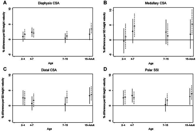
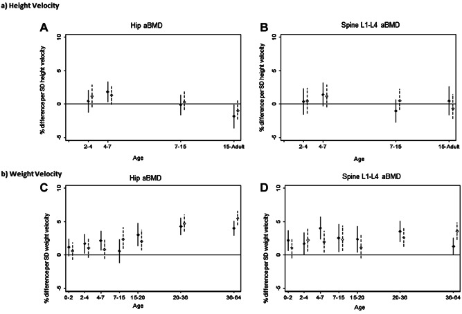
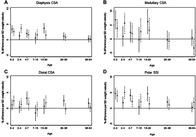
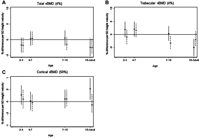
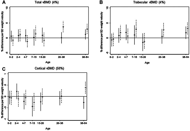
There is growing evidence that early growth influences bone mass in later life but most studies are limited to birth weight and/or early infant growth and dual-energy X-ray absorptiometry (DXA) measurements. In a British birth cohort study with prospective measures of lifetime height and weight, we investigated the growth trajectory in relation to bone in males (M) and females (F) at 60 to 64 years old. Outcomes were DXA measures of hip and spine areal bone density (aBMD) (n = 1658) and pQCT measures of distal and diaphyseal radius cross-sectional area (CSA), strength, and volumetric bone density (vBMD) (n = 1350 of the 1658). Regression models examined percentage change in bone parameters with standardized measures of birth weight, height, and weight. A series of conditional growth models were fitted for height and weight gain (using intervals: birth-2, 2-4, 4-7, 7-15, 15-20, 20-36, and 36-64 years) and height gain (using intervals: 2-4, 4-7, 7-15, and 15-36 years). Birth weight was positively related to bone CSA (M: 1.4%; 95% confidence interval [CI], 0.3%-2.5%; F: 1.3%; 95% CI, 0.3%-2.4% per 1 SD increase in birth weight for diaphyseal CSA) and strength (M: 1.8%; 95% CI, 0.3-3.4; F: 2.0%; 95% CI, 0.5-3.5). No positive associations were found with trabecular, total, or cortical vBMD. One SD change in prepubertal and postpubertal height and weight velocities were associated with between 2% and 5% greater bone CSA and strength. Height gain in later years was negatively associated with trabecular vBMD. Weight gain velocity during the adult years was positively associated with up to 4% greater trabecular and total BMD, and 4% greater aBMD at hip and spine. In a cohort born in the early post-war period, higher birth weight, gaining weight and height faster than others, particularly through the prepubertal and postpubertal periods, was positively related to bone strength, mostly through greater bone CSA, at 60 to 64 years.
越来越多的证据表明,早期生长会影响晚年的骨量,但大多数研究仅限于出生体重和/或婴儿早期生长以及双能X线吸收法(DXA)测量。在一项对身高和体重进行前瞻性测量的英国出生队列研究中,我们调查了60至64岁男性(M)和女性(F)的生长轨迹与骨骼的关系。结局指标为DXA测量的髋部和脊柱面积骨密度(aBMD)(n = 1658)以及pQCT测量的桡骨远端和骨干的横截面积(CSA)、强度和体积骨密度(vBMD)(1658例中的1350例)。回归模型用出生体重、身高和体重的标准化测量值来检验骨骼参数的百分比变化。针对身高和体重增加(使用区间:出生至2岁、2至4岁、4至7岁、7至15岁、15至20岁、20至36岁以及36至64岁)以及身高增加(使用区间:2至4岁、4至7岁、7至15岁以及15至36岁)拟合了一系列条件生长模型。出生体重与骨骼CSA呈正相关(骨干CSA:男性为1.4%;95%置信区间[CI],0.3% - 2.5%;女性为1.3%;95%CI,0.3% - 2.4%,出生体重每增加1个标准差)以及强度(男性为1.8%;95%CI,0.3 - 3.4;女性为2.0%;95%CI,0.5 - 3.5)。未发现与小梁、总体或皮质vBMD有正相关。青春期前和青春期后的身高和体重速度每变化1个标准差,骨骼CSA和强度会增加2%至5%。晚年的身高增加与小梁vBMD呈负相关。成年期的体重增加速度与小梁和总体BMD增加高达4%以及髋部和脊柱aBMD增加4%呈正相关。在战后早期出生的队列中,较高的出生体重、比其他人更快地增加体重和身高,尤其是在青春期前和青春期后阶段,在60至64岁时与骨骼强度呈正相关,主要是通过更大的骨骼CSA实现的。