• 文献检索
  • 文档翻译
  • 深度研究
  • 学术资讯
  • Suppr Zotero 插件Zotero 插件
  • 邀请有礼
  • 套餐&价格
  • 历史记录
应用&插件
Suppr Zotero 插件Zotero 插件浏览器插件Mac 客户端Windows 客户端微信小程序
定价
高级版会员购买积分包购买API积分包
服务
文献检索文档翻译深度研究API 文档MCP 服务
关于我们
关于 Suppr公司介绍联系我们用户协议隐私条款
关注我们

Suppr 超能文献

核心技术专利:CN118964589B侵权必究
粤ICP备2023148730 号-1Suppr @ 2026

文献检索

告别复杂PubMed语法,用中文像聊天一样搜索,搜遍4000万医学文献。AI智能推荐,让科研检索更轻松。

立即免费搜索

文件翻译

保留排版,准确专业,支持PDF/Word/PPT等文件格式,支持 12+语言互译。

免费翻译文档

深度研究

AI帮你快速写综述,25分钟生成高质量综述,智能提取关键信息,辅助科研写作。

立即免费体验

面神经成像:当代综述

Imaging the facial nerve: a contemporary review.

作者信息

Gupta Sachin, Mends Francine, Hagiwara Mari, Fatterpekar Girish, Roehm Pamela C

机构信息

Department of Otolaryngology, New York University School of Medicine, New York, NY 10016, USA.

出版信息

Radiol Res Pract. 2013;2013:248039. doi: 10.1155/2013/248039. Epub 2013 May 23.

DOI:10.1155/2013/248039
PMID:23766904
原文链接:https://pmc.ncbi.nlm.nih.gov/articles/PMC3676972/
Abstract

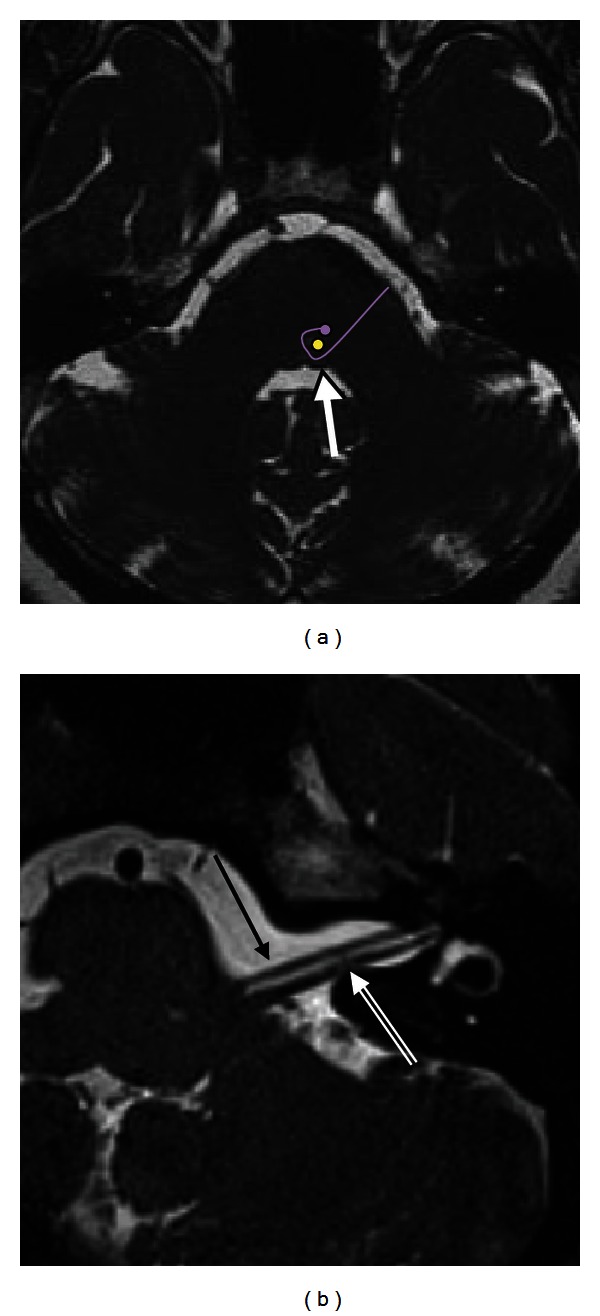
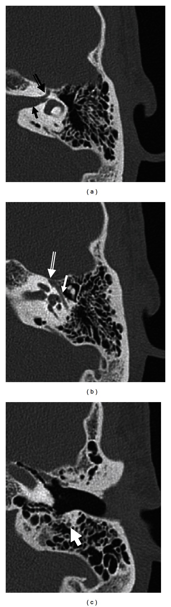
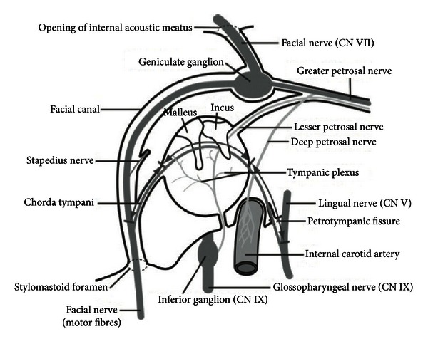
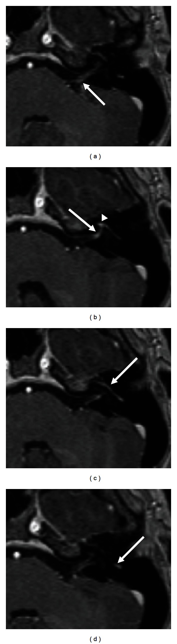
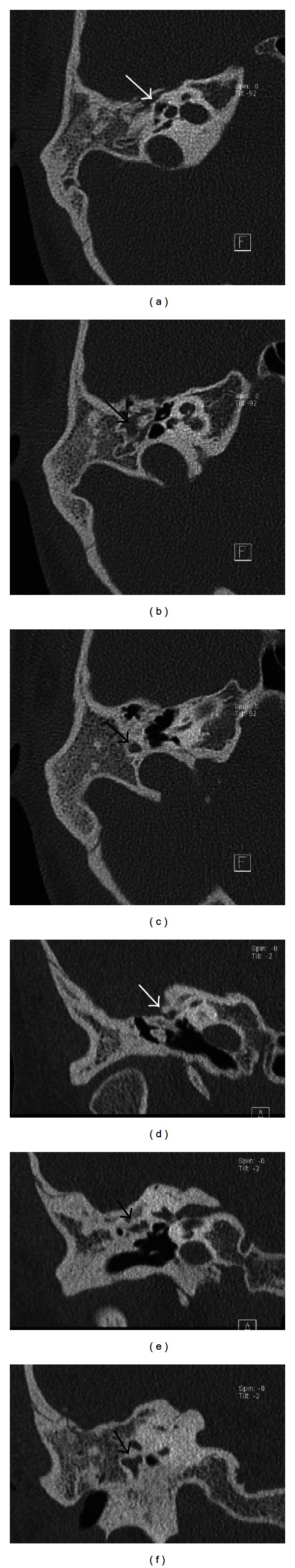
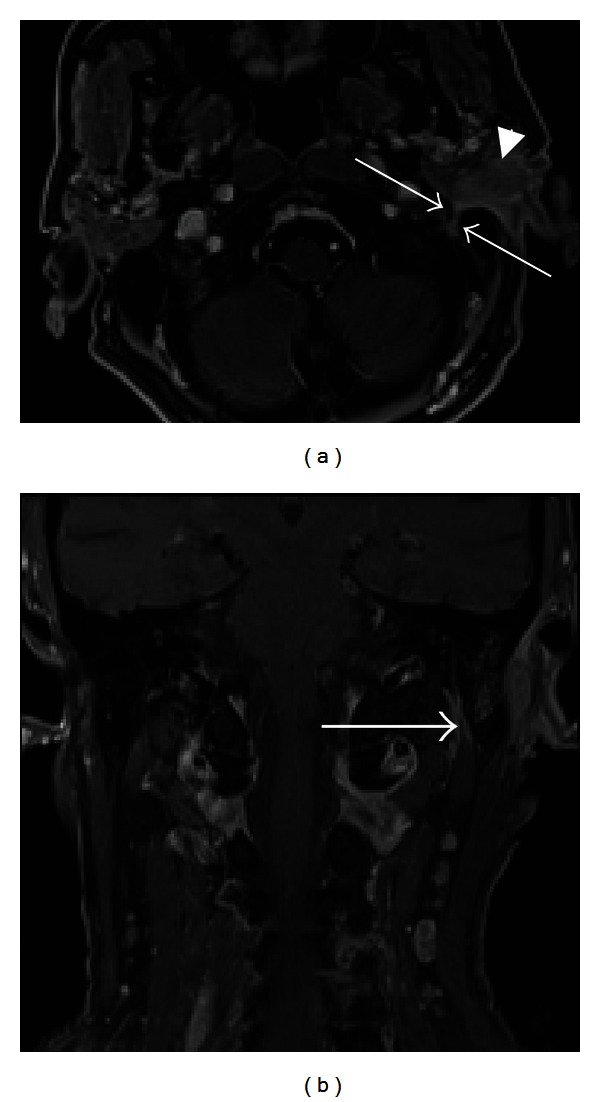
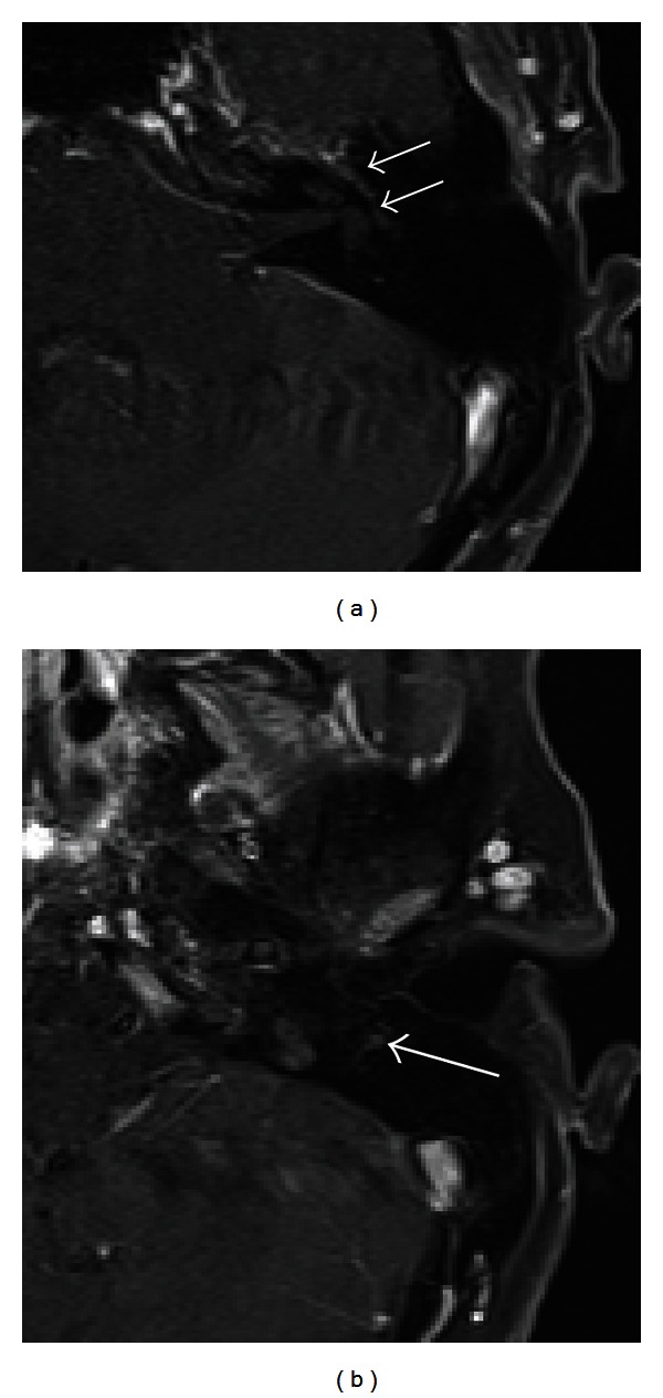
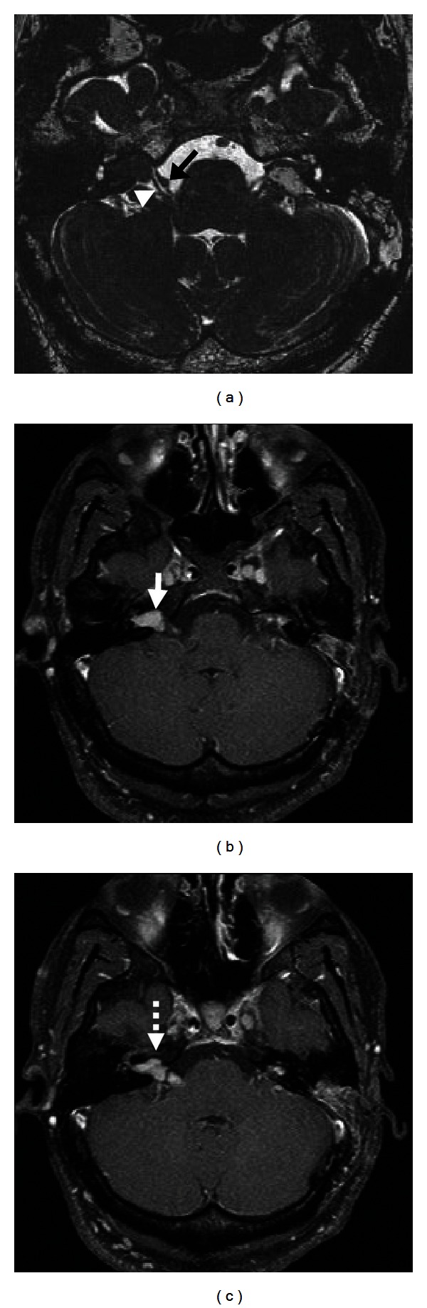
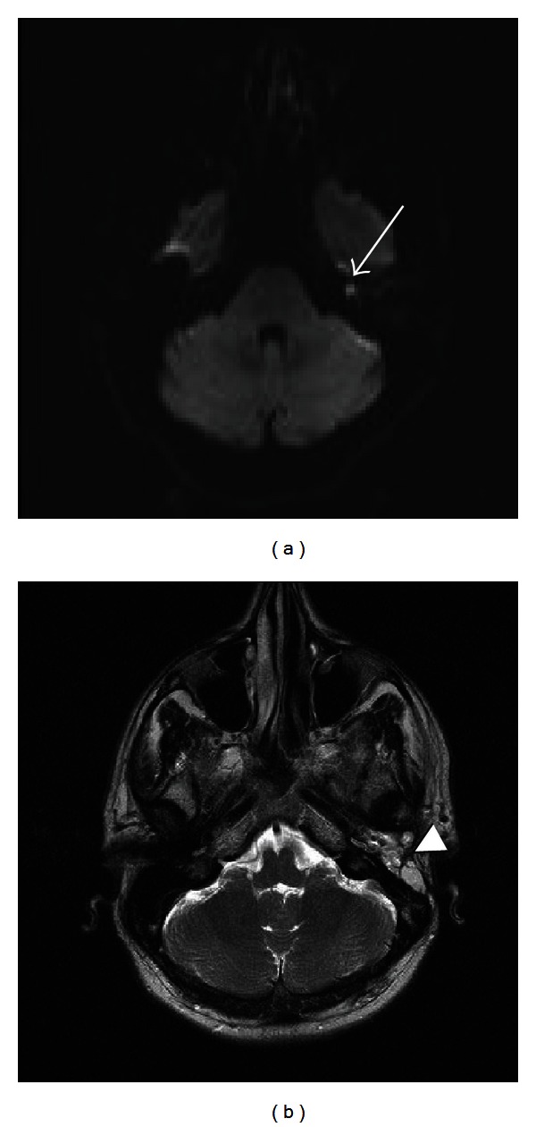
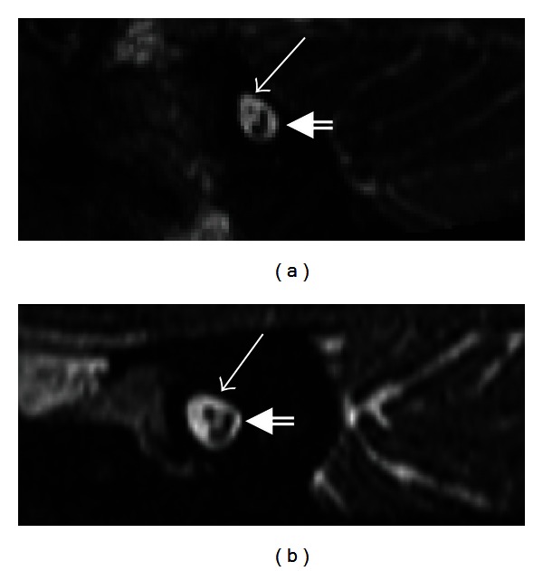
Imaging plays a critical role in the evaluation of a number of facial nerve disorders. The facial nerve has a complex anatomical course; thus, a thorough understanding of the course of the facial nerve is essential to localize the sites of pathology. Facial nerve dysfunction can occur from a variety of causes, which can often be identified on imaging. Computed tomography and magnetic resonance imaging are helpful for identifying bony facial canal and soft tissue abnormalities, respectively. Ultrasound of the facial nerve has been used to predict functional outcomes in patients with Bell's palsy. More recently, diffusion tensor tractography has appeared as a new modality which allows three-dimensional display of facial nerve fibers.

摘要

影像学在多种面神经疾病的评估中起着关键作用。面神经具有复杂的解剖走行;因此,对面神经走行的透彻理解对于定位病变部位至关重要。面神经功能障碍可由多种原因引起,这些原因通常可通过影像学检查识别。计算机断层扫描和磁共振成像分别有助于识别面神经骨管和软组织异常。面神经超声已被用于预测贝尔面瘫患者的功能预后。最近,扩散张量纤维束成像作为一种新的检查方法出现,它能够三维显示面神经纤维。

https://cdn.ncbi.nlm.nih.gov/pmc/blobs/4df9/3676972/bc26e312ccd3/RRP2013-248039.015.jpg
https://cdn.ncbi.nlm.nih.gov/pmc/blobs/4df9/3676972/694689ce25c8/RRP2013-248039.001.jpg
https://cdn.ncbi.nlm.nih.gov/pmc/blobs/4df9/3676972/455e0fee5792/RRP2013-248039.002.jpg
https://cdn.ncbi.nlm.nih.gov/pmc/blobs/4df9/3676972/ff490a5ce8d1/RRP2013-248039.003.jpg
https://cdn.ncbi.nlm.nih.gov/pmc/blobs/4df9/3676972/d24d09036a58/RRP2013-248039.004.jpg
https://cdn.ncbi.nlm.nih.gov/pmc/blobs/4df9/3676972/f86afff424a6/RRP2013-248039.005.jpg
https://cdn.ncbi.nlm.nih.gov/pmc/blobs/4df9/3676972/d832247d85b9/RRP2013-248039.006.jpg
https://cdn.ncbi.nlm.nih.gov/pmc/blobs/4df9/3676972/329926aad058/RRP2013-248039.007.jpg
https://cdn.ncbi.nlm.nih.gov/pmc/blobs/4df9/3676972/223dabc23235/RRP2013-248039.008.jpg
https://cdn.ncbi.nlm.nih.gov/pmc/blobs/4df9/3676972/db85bd9438f5/RRP2013-248039.009.jpg
https://cdn.ncbi.nlm.nih.gov/pmc/blobs/4df9/3676972/350312d6c065/RRP2013-248039.010.jpg
https://cdn.ncbi.nlm.nih.gov/pmc/blobs/4df9/3676972/303478328926/RRP2013-248039.011.jpg
https://cdn.ncbi.nlm.nih.gov/pmc/blobs/4df9/3676972/814f2224ee31/RRP2013-248039.012.jpg
https://cdn.ncbi.nlm.nih.gov/pmc/blobs/4df9/3676972/e5e23320c3d4/RRP2013-248039.013.jpg
https://cdn.ncbi.nlm.nih.gov/pmc/blobs/4df9/3676972/a6555a5a3cd4/RRP2013-248039.014.jpg
https://cdn.ncbi.nlm.nih.gov/pmc/blobs/4df9/3676972/bc26e312ccd3/RRP2013-248039.015.jpg

相似文献

1
Imaging the facial nerve: a contemporary review.面神经成像:当代综述
Radiol Res Pract. 2013;2013:248039. doi: 10.1155/2013/248039. Epub 2013 May 23.
2
The ratio of facial nerve to facial canal as an indicator of entrapment in Bell's palsy: A study by CT and MRI.面神经与面神经管比值作为 Bell 麻痹时神经受压的指标:CT 和 MRI 研究。
Clin Neurol Neurosurg. 2020 Nov;198:106109. doi: 10.1016/j.clineuro.2020.106109. Epub 2020 Jul 25.
3
The facial nerve canal in patients with Bell's palsy: an investigation by high-resolution computed tomography with multiplanar reconstruction.贝尔氏麻痹症患者的面神经管:高分辨率 CT 多平面重建的研究。
Eur Arch Otorhinolaryngol. 2013 Jul;270(7):2035-8. doi: 10.1007/s00405-012-2253-7. Epub 2012 Nov 11.
4
Facial palsy from temporal bone lesions.颞骨病变导致的面瘫。
Ann Acad Med Singap. 2005 May;34(4):322-9.
5
Clinical practice guideline: Bell's palsy.临床实践指南:贝尔氏麻痹。
Otolaryngol Head Neck Surg. 2013 Nov;149(3 Suppl):S1-27. doi: 10.1177/0194599813505967.
6
[Electrophysiological study on pathophysiology of Bell's palsy--distribution of nerve conduction velocities (DNCV) in facial nerve with collision method].[贝尔麻痹病理生理学的电生理研究——采用碰撞法对面神经神经传导速度分布(DNCV)的研究]
Nihon Jibiinkoka Gakkai Kaiho. 1991 Jul;94(7):906-14. doi: 10.3950/jibiinkoka.94.906.
7
Contrast-enhanced MR imaging of the facial nerve in 11 patients with Bell's palsy.11例贝尔面瘫患者面神经的对比增强磁共振成像
AJR Am J Roentgenol. 1990 Sep;155(3):573-9. doi: 10.2214/ajr.155.3.2117359.
8
Contrast-enhanced MR imaging of the facial nerve in 11 patients with Bell's palsy.11例贝尔麻痹患者面神经的对比增强磁共振成像
AJNR Am J Neuroradiol. 1990 Jul-Aug;11(4):735-41.
9
Measurement of the depth of facial nerve at the level of stylomastoid foramen using MR imaging in Bell's palsy.磁共振成像测量贝尔面瘫患者茎乳孔水平面神经深度。
Clin Imaging. 2019 Nov-Dec;58:34-38. doi: 10.1016/j.clinimag.2019.06.008. Epub 2019 Jun 14.
10
Is the width of the labyrinthine portion of the fallopian tube implicated in the pathophysiology of Bell's palsy?: a prospective clinical study using computed tomography.输卵管迷路部的宽度与贝尔氏面瘫的病理生理学有关吗?:一项使用计算机断层扫描的前瞻性临床研究。
Laryngoscope. 2010 Jun;120(6):1203-7. doi: 10.1002/lary.20896.

引用本文的文献

1
3D T1-Weighted Black-Blood MRI in the Diagnosis and Follow-Up of Facial Neuritis: a Single-Center Prospective Study.3D T1加权黑血磁共振成像在面神经炎诊断及随访中的应用:一项单中心前瞻性研究
Clin Neuroradiol. 2025 Jul 14. doi: 10.1007/s00062-025-01540-5.
2
An Interdisciplinary Review of the Zygomaticus Muscles: Anatomical Variability, Imaging Modalities, and Clinical Implications.颧肌的跨学科综述:解剖变异性、成像方式及临床意义
J Clin Med. 2025 Jun 10;14(12):4110. doi: 10.3390/jcm14124110.
3
Abnormalities of the Facial Nerve in Temporal Bones With Inner Ear Malformations.

本文引用的文献

1
Neuroimaging in acute stroke.急性卒中的神经影像学
Emerg Med Clin North Am. 2012 Aug;30(3):659-80. doi: 10.1016/j.emc.2012.06.005.
2
Facial nerve hemangioma: a rare case involving the vertical segment.面神经血管瘤:累及垂直段的罕见病例。
Laryngoscope. 2013 Feb;123(2):499-502. doi: 10.1002/lary.23431. Epub 2012 Jul 9.
3
Enlarged geniculate ganglion fossa: CT sign of facial nerve canal fracture.镫骨神经节窝扩大:面神经管骨折的 CT 征象。
内耳畸形颞骨中面神经的异常
Laryngoscope Investig Otolaryngol. 2025 Apr 22;10(2):e70146. doi: 10.1002/lio2.70146. eCollection 2025 Apr.
4
Rare case of aberrant course of facial nerve traversing above oval window.面神经走行异常越过椭圆窗的罕见病例。
Radiol Case Rep. 2025 Mar 17;20(6):2794-2796. doi: 10.1016/j.radcr.2024.09.101. eCollection 2025 Jun.
5
Double echo steady-state free precession technique in MR neurography to evaluate extracranial facial nerve involvement in a case of Bell's palsy.磁共振神经成像中双回波稳态自由进动技术用于评估贝尔麻痹病例中颅外面神经受累情况。
Radiol Case Rep. 2024 Aug 14;19(11):4904-4907. doi: 10.1016/j.radcr.2024.07.130. eCollection 2024 Nov.
6
Stapedotomy for Pediatric Middle Ear Anomalies With Facial Nerve Bifurcation: A Case Report.面神经分叉处小儿中耳畸形的镫骨切除术:一例报告
Cureus. 2024 Jul 25;16(7):e65391. doi: 10.7759/cureus.65391. eCollection 2024 Jul.
7
Determination of the radioanatomical factors related to the position of facial nerve in accessing jugular foramen and carotid artery in temporal bone.颞骨中与面神经在进入颈静脉孔和颈动脉位置相关的放射解剖学因素的确定。
Heliyon. 2024 Feb 16;10(4):e26560. doi: 10.1016/j.heliyon.2024.e26560. eCollection 2024 Feb 29.
8
Variability of the Cervical Branch Depending on the Facial Nerve Branching Pattern and Anthropometric Type of the Head.根据面神经分支模式和头部人体测量类型,颈支的变异性
Neurol Int. 2024 Jan 4;16(1):113-125. doi: 10.3390/neurolint16010007.
9
Labyrinthine Bifurcation of the Facial Nerve.面神经的迷路分叉
Indian J Otolaryngol Head Neck Surg. 2023 Dec;75(4):4086-4089. doi: 10.1007/s12070-023-03977-x. Epub 2023 Jul 15.
10
Intra-parotid facial nerve path by MRI tractography: radio-clinical comparison in parotid tumors.MRI 轨迹法显示腮腺内面神经径路:腮腺肿瘤的放射-临床对照研究。
Eur Arch Otorhinolaryngol. 2024 Feb;281(2):925-934. doi: 10.1007/s00405-023-08301-5. Epub 2023 Nov 2.
Acad Radiol. 2012 Aug;19(8):971-6. doi: 10.1016/j.acra.2012.03.025.
4
Dissection and exposure of the whole course of deep nerves in human head specimens after decalcification.脱钙后人头颅标本中深部神经全程的解剖与暴露
Int J Otolaryngol. 2012;2012:418650. doi: 10.1155/2012/418650. Epub 2012 Mar 15.
5
Complications of chronic suppurative otitis media: a retrospective review.慢性化脓性中耳炎并发症:回顾性研究。
Eur Arch Otorhinolaryngol. 2013 Jan;270(1):69-76. doi: 10.1007/s00405-012-1924-8. Epub 2012 Jan 15.
6
Paraganglioma of the facial nerve, a rare differential diagnosis for facial nerve paralysis: case report and review of the literature.面神经副神经节瘤,面神经麻痹的罕见鉴别诊断:病例报告及文献复习。
Eur Arch Otorhinolaryngol. 2012 Feb;269(2):693-8. doi: 10.1007/s00405-011-1840-3. Epub 2011 Nov 20.
7
Normal and pathological findings for the facial nerve on magnetic resonance imaging.磁共振成像对面神经正常和病理表现的评估。
Clin Radiol. 2011 Aug;66(8):701-7. doi: 10.1016/j.crad.2011.02.012. Epub 2011 Apr 22.
8
Three-dimensional in vivo modeling of vestibular schwannomas and surrounding cranial nerves with diffusion imaging tractography.应用弥散张量成像技术对前庭神经鞘瘤及其周围颅神经进行三维活体建模。
Neurosurgery. 2011 Apr;68(4):1077-83. doi: 10.1227/NEU.0b013e31820c6cbe.
9
Development of the stapedius muscle canal and its possible clinical consequences.镫骨肌管的发育及其可能的临床后果。
Int J Pediatr Otorhinolaryngol. 2011 Feb;75(2):277-81. doi: 10.1016/j.ijporl.2010.11.018. Epub 2010 Dec 10.
10
Prospective blinded study of computed tomography in congenital aural atresia.先天性外耳道闭锁计算机断层扫描的前瞻性盲法研究。
Int J Pediatr Otorhinolaryngol. 2010 Nov;74(11):1286-91. doi: 10.1016/j.ijporl.2010.08.006.