• 文献检索
  • 文档翻译
  • 深度研究
  • 学术资讯
  • Suppr Zotero 插件Zotero 插件
  • 邀请有礼
  • 套餐&价格
  • 历史记录
应用&插件
Suppr Zotero 插件Zotero 插件浏览器插件Mac 客户端Windows 客户端微信小程序
定价
高级版会员购买积分包购买API积分包
服务
文献检索文档翻译深度研究API 文档MCP 服务
关于我们
关于 Suppr公司介绍联系我们用户协议隐私条款
关注我们

Suppr 超能文献

核心技术专利:CN118964589B侵权必究
粤ICP备2023148730 号-1Suppr @ 2026

文献检索

告别复杂PubMed语法,用中文像聊天一样搜索,搜遍4000万医学文献。AI智能推荐,让科研检索更轻松。

立即免费搜索

文件翻译

保留排版,准确专业,支持PDF/Word/PPT等文件格式,支持 12+语言互译。

免费翻译文档

深度研究

AI帮你快速写综述,25分钟生成高质量综述,智能提取关键信息,辅助科研写作。

立即免费体验

使用高分辨率μCT对皮质骨孔隙率和组织矿物质密度进行三维评估:分辨率和阈值方法的影响

3D assessment of cortical bone porosity and tissue mineral density using high-resolution µCT: effects of resolution and threshold method.

作者信息

Palacio-Mancheno Paolo E, Larriera Adriana I, Doty Stephen B, Cardoso Luis, Fritton Susannah P

机构信息

Department of Biomedical Engineering, City College of New York, New York, NY, USA.

出版信息

J Bone Miner Res. 2014 Jan;29(1):142-50. doi: 10.1002/jbmr.2012.

DOI:10.1002/jbmr.2012
PMID:23775635
原文链接:https://pmc.ncbi.nlm.nih.gov/articles/PMC3870034/
Abstract

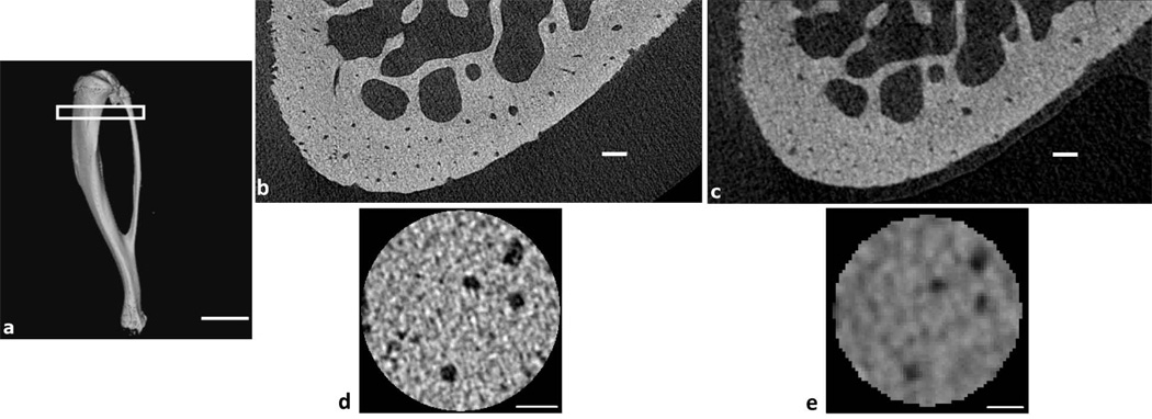
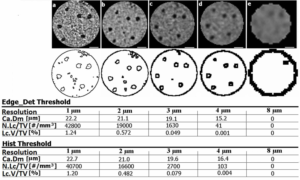
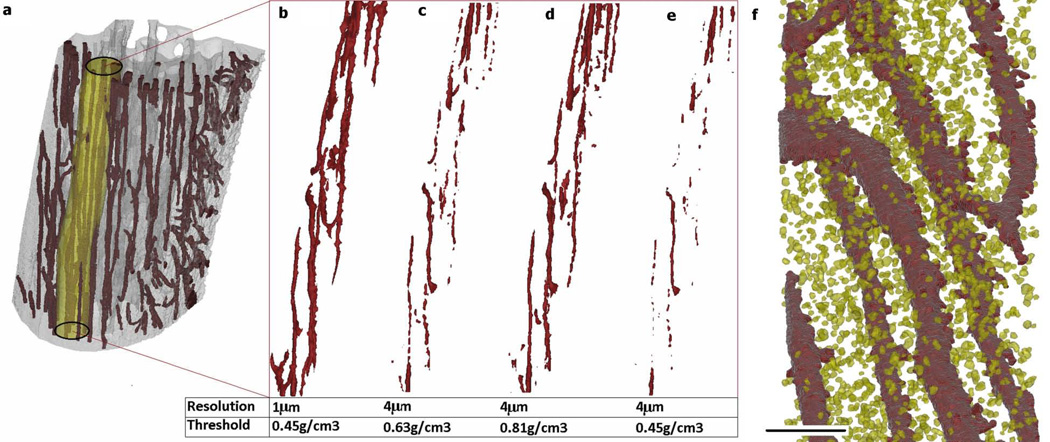
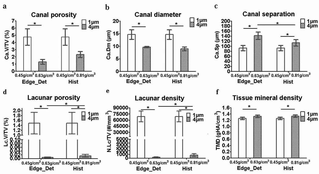
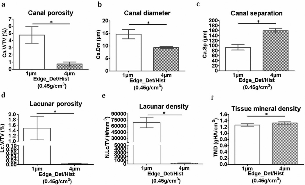
Current micro-computed tomography (µCT) systems allow scanning bone at resolutions capable of three-dimensional (3D) characterization of intracortical vascular porosity and osteocyte lacunae. However, the scanning and reconstruction parameters along with the image segmentation method affect the accuracy of the measurements. In this study, the effects of scanning resolution and image threshold method in quantifying small features of cortical bone (vascular porosity, vascular canal diameter and separation, lacunar porosity and density, and tissue mineral density) were analyzed. Cortical bone from the tibia of Sprague-Dawley rats was scanned at 1-µm and 4-µm resolution, reconstructions were density-calibrated, and volumes of interest were segmented using approaches based on edge-detection or histogram analysis. In 1-µm resolution scans, the osteocyte lacunar spaces could be visualized, and it was possible to separate the lacunar porosity from the vascular porosity. At 4-µm resolution, the vascular porosity and vascular canal diameter were underestimated, and osteocyte lacunae were not effectively detected, whereas the vascular canal separation and tissue mineral density were overestimated compared to 1-µm resolution. Resolution had a much greater effect on the measurements than did threshold method, showing partial volume effects at resolutions coarser than 2 µm in two separate analyses, one of which assessed the effect of resolution on an object of known size with similar architecture to a vascular pore. Although there was little difference when using the edge-detection versus histogram-based threshold approaches, edge-detection was somewhat more effective in delineating canal architecture at finer resolutions (1-2 µm). In addition, use of a high-resolution (1 µm) density-based threshold on lower resolution (4 µm) density-calibrated images was not effective in improving the lower-resolution measurements. In conclusion, if measuring cortical vascular microarchitecture, especially in small animals, a µCT resolution of 1 to 2 µm is appropriate, whereas a resolution of at least 1 µm is necessary when assessing osteocyte lacunar porosity.

摘要

当前的微型计算机断层扫描(µCT)系统能够以三维(3D)方式对皮质内血管孔隙率和骨细胞陷窝进行表征的分辨率来扫描骨骼。然而,扫描和重建参数以及图像分割方法会影响测量的准确性。在本研究中,分析了扫描分辨率和图像阈值方法对量化皮质骨小特征(血管孔隙率、血管管径和间距、陷窝孔隙率和密度以及组织矿物质密度)的影响。对来自Sprague-Dawley大鼠胫骨的皮质骨以1微米和4微米的分辨率进行扫描,对重建图像进行密度校准,并使用基于边缘检测或直方图分析的方法对感兴趣区域进行分割。在1微米分辨率扫描中,可以观察到骨细胞陷窝空间,并且能够将陷窝孔隙率与血管孔隙率区分开来。在4微米分辨率下,血管孔隙率和血管管径被低估,骨细胞陷窝未被有效检测到,而与1微米分辨率相比,血管管径间距和组织矿物质密度被高估。分辨率对测量的影响比阈值方法大得多,在两项独立分析中,在大于2微米的分辨率下显示出部分容积效应,其中一项分析评估了分辨率对具有与血管孔相似结构的已知大小物体的影响。尽管使用基于边缘检测和基于直方图的阈值方法时差异不大,但在更精细的分辨率(1至2微米)下,边缘检测在描绘管道结构方面更有效。此外,在较低分辨率(4微米)的密度校准图像上使用基于高分辨率(1微米)密度的阈值并不能有效改善较低分辨率的测量结果。总之,如果要测量皮质血管微结构,尤其是在小动物中,µCT分辨率为1至2微米是合适的,而在评估骨细胞陷窝孔隙率时,至少1微米的分辨率是必要的。

https://cdn.ncbi.nlm.nih.gov/pmc/blobs/c76f/3870034/63fa8719ea40/nihms497828f6.jpg
https://cdn.ncbi.nlm.nih.gov/pmc/blobs/c76f/3870034/3cb13d3b7fd8/nihms497828f1.jpg
https://cdn.ncbi.nlm.nih.gov/pmc/blobs/c76f/3870034/389638b5888f/nihms497828f2.jpg
https://cdn.ncbi.nlm.nih.gov/pmc/blobs/c76f/3870034/2abcdce1a8b4/nihms497828f3.jpg
https://cdn.ncbi.nlm.nih.gov/pmc/blobs/c76f/3870034/5cad12bf0a06/nihms497828f4.jpg
https://cdn.ncbi.nlm.nih.gov/pmc/blobs/c76f/3870034/cedac88d3ff3/nihms497828f5.jpg
https://cdn.ncbi.nlm.nih.gov/pmc/blobs/c76f/3870034/63fa8719ea40/nihms497828f6.jpg

相似文献

1
3D assessment of cortical bone porosity and tissue mineral density using high-resolution µCT: effects of resolution and threshold method.使用高分辨率μCT对皮质骨孔隙率和组织矿物质密度进行三维评估:分辨率和阈值方法的影响
J Bone Miner Res. 2014 Jan;29(1):142-50. doi: 10.1002/jbmr.2012.
2
3D visualization and quantification of rat cortical bone porosity using a desktop micro-CT system: a case study in the tibia.使用桌面型微计算机断层扫描系统对大鼠皮质骨孔隙率进行 3D 可视化和定量分析:以胫骨为例的研究。
J Microsc. 2010 Oct;240(1):32-7. doi: 10.1111/j.1365-2818.2010.03381.x.
3
Multiscale and multimodality computed tomography for cortical bone analysis.用于皮质骨分析的多尺度和多模态计算机断层扫描
Phys Med Biol. 2016 Dec 21;61(24):8553-8576. doi: 10.1088/0031-9155/61/24/8553. Epub 2016 Nov 15.
4
Botox-induced muscle paralysis alters intracortical porosity and osteocyte lacunar density in skeletally mature rats.肉毒杆菌毒素诱导的肌肉瘫痪改变成熟骨骼大鼠皮质内孔隙率和骨细胞陷窝密度。
J Orthop Res. 2019 May;37(5):1153-1163. doi: 10.1002/jor.24276. Epub 2019 Mar 28.
5
The effects of estrogen deficiency on cortical bone microporosity and mineralization.雌激素缺乏对皮质骨微孔结构和矿化的影响。
Bone. 2018 May;110:1-10. doi: 10.1016/j.bone.2018.01.019. Epub 2018 Jan 31.
6
Interstitial fluid velocity is decreased around cortical bone vascular pores and depends on osteocyte position in a rat model of disuse osteoporosis.在废用性骨质疏松症大鼠模型中,皮质骨血管孔周围的间质液速度降低,且依赖于骨细胞的位置。
Biomech Model Mechanobiol. 2021 Jun;20(3):1135-1146. doi: 10.1007/s10237-021-01438-4. Epub 2021 Mar 5.
7
Alterations in the osteocyte lacunar-canalicular microenvironment due to estrogen deficiency.由于雌激素缺乏导致的破骨细胞陷窝-小管微环境的改变。
Bone. 2012 Sep;51(3):488-97. doi: 10.1016/j.bone.2012.05.014. Epub 2012 May 23.
8
Large-scale quantification of human osteocyte lacunar morphological biomarkers as assessed by ultra-high-resolution desktop micro-computed tomography.采用超高分辨率台式微计算机断层扫描技术对人成骨细胞陷窝形态生物标志物进行大规模定量分析。
Bone. 2021 Nov;152:116094. doi: 10.1016/j.bone.2021.116094. Epub 2021 Jun 27.
9
3D osteocyte lacunar morphometric properties and distributions in human femoral cortical bone using synchrotron radiation micro-CT images.利用同步辐射微计算机断层扫描图像研究人股骨皮质骨中 3D 骨细胞陷窝的形态计量学特征及其分布
Bone. 2014 Mar;60:172-85. doi: 10.1016/j.bone.2013.12.008. Epub 2013 Dec 12.
10
Ultrastructural properties in cortical bone vary greatly in two inbred strains of mice as assessed by synchrotron light based micro- and nano-CT.通过基于同步加速器光的微米和纳米CT评估,两种近交系小鼠的皮质骨超微结构特性差异很大。
J Bone Miner Res. 2007 Oct;22(10):1557-70. doi: 10.1359/jbmr.070703.

引用本文的文献

1
In Vivo Assessment of a Triple Periodic Minimal Surface Based Biomimmetic Gyroid as an Implant Material in a Rabbit Tibia Model.基于三重周期极小曲面的仿生类螺旋体作为植入材料在兔胫骨模型中的体内评估
ACS Mater Au. 2024 Jun 13;4(5):479-488. doi: 10.1021/acsmaterialsau.4c00016. eCollection 2024 Sep 11.
2
Influence of micro-structural parameters on apparent absorption coefficient in porous structures mimicking cortical bone.微观结构参数对模拟皮质骨的多孔结构中表观吸收系数的影响。
IEEE Int Ultrason Symp. 2018 Oct;2018. doi: 10.1109/ultsym.2018.8579610. Epub 2018 Dec 20.
3
2D vs. 3D Evaluation of Osteocyte Lacunae - Methodological Approaches, Recommended Parameters, and Challenges: A Narrative Review by the European Calcified Tissue Society (ECTS).

本文引用的文献

1
Advances in assessment of bone porosity, permeability and interstitial fluid flow.骨孔隙率、渗透性和细胞间液流动评估的进展。
J Biomech. 2013 Jan 18;46(2):253-65. doi: 10.1016/j.jbiomech.2012.10.025. Epub 2012 Nov 19.
2
High resolution micro arthrography of hard and soft tissues in a murine model.高分辨率微关节造影术在小鼠模型中的硬组织和软组织成像。
Osteoarthritis Cartilage. 2012 Sep;20(9):1011-9. doi: 10.1016/j.joca.2012.05.004. Epub 2012 May 18.
3
Changes in intracortical microporosities induced by pharmaceutical treatment of osteoporosis as detected by high resolution micro-CT.
2D 与 3D 评估破骨细胞陷窝:方法学方法、推荐参数和挑战:欧洲钙化组织学会 (ECTS) 的叙述性综述。
Curr Osteoporos Rep. 2024 Aug;22(4):396-415. doi: 10.1007/s11914-024-00877-z. Epub 2024 Jul 9.
4
Computed tomography technologies to measure key structural features of polymeric biomedical implants from bench to bedside.从实验室到临床,计算断层扫描技术可用于测量聚合物生物医学植入物的关键结构特征。
J Biomed Mater Res A. 2024 Nov;112(11):1893-1901. doi: 10.1002/jbm.a.37735. Epub 2024 May 10.
5
Deep learning to overcome Zernike phase-contrast nanoCT artifacts for automated micro-nano porosity segmentation in bone.深度学习克服泽尼克相衬纳米 CT 伪影,实现骨微纳米孔隙率的自动分割。
J Synchrotron Radiat. 2024 Jan 1;31(Pt 1):136-149. doi: 10.1107/S1600577523009852.
6
Sericin/Nano-Hydroxyapatite Hydrogels Based on Graphene Oxide for Effective Bone Regeneration via Immunomodulation and Osteoinduction.基于氧化石墨烯的丝胶/纳米羟基磷灰石水凝胶通过免疫调节和成骨诱导实现有效的骨再生。
Int J Nanomedicine. 2023 Apr 6;18:1875-1895. doi: 10.2147/IJN.S399487. eCollection 2023.
7
In Vitro and In Vivo Biocompatible and Controlled Resveratrol Release Performances of HEMA/Alginate and HEMA/Gelatin IPN Hydrogel Scaffolds.HEMA/藻酸盐和HEMA/明胶互穿网络水凝胶支架的体外和体内生物相容性及白藜芦醇可控释放性能
Polymers (Basel). 2022 Oct 21;14(20):4459. doi: 10.3390/polym14204459.
8
Therapeutic Targeting Notch2 Protects Bone Micro-Vasculatures from Methotrexate Chemotherapy-Induced Adverse Effects in Rats.靶向治疗 Notch2 可保护大鼠的骨微血管免受甲氨蝶呤化疗的不良影响。
Cells. 2022 Aug 2;11(15):2382. doi: 10.3390/cells11152382.
9
Comparison of the 3D-Microstructure Between Alveolar and Iliac Bone for Enhanced Bioinspired Bone Graft Substitutes.用于增强仿生骨移植替代物的牙槽骨和髂骨三维微观结构比较
Front Bioeng Biotechnol. 2022 Jun 17;10:862395. doi: 10.3389/fbioe.2022.862395. eCollection 2022.
10
Temporal changes in cortical microporosity during estrogen deficiency associated with perilacunar resorption and osteocyte apoptosis: A pilot study.雌激素缺乏相关的腔隙周围吸收和骨细胞凋亡过程中皮质微孔隙度的时间变化:一项初步研究。
Bone Rep. 2022 May 17;16:101590. doi: 10.1016/j.bonr.2022.101590. eCollection 2022 Jun.
高分辨率微计算机断层扫描检测骨质疏松症药物治疗引起的皮质内微孔变化。
Bone. 2012 Mar;50(3):596-604. doi: 10.1016/j.bone.2011.12.012. Epub 2011 Dec 28.
4
3D visualization and quantification of rat cortical bone porosity using a desktop micro-CT system: a case study in the tibia.使用桌面型微计算机断层扫描系统对大鼠皮质骨孔隙率进行 3D 可视化和定量分析:以胫骨为例的研究。
J Microsc. 2010 Oct;240(1):32-7. doi: 10.1111/j.1365-2818.2010.03381.x.
5
Effects of CT image segmentation methods on the accuracy of long bone 3D reconstructions.CT 图像分割方法对长骨 3D 重建准确性的影响。
Med Eng Phys. 2011 Mar;33(2):226-33. doi: 10.1016/j.medengphy.2010.10.002. Epub 2010 Oct 27.
6
Guidelines for assessment of bone microstructure in rodents using micro-computed tomography.骨组织形态计量学分析的鼠类骨骼microCT 评估指南
J Bone Miner Res. 2010 Jul;25(7):1468-86. doi: 10.1002/jbmr.141.
7
Hierarchical microimaging of bone structure and function.骨结构与功能的分层显微成像
Nat Rev Rheumatol. 2009 Jul;5(7):373-81. doi: 10.1038/nrrheum.2009.107.
8
Simultaneous 3D visualization and quantification of murine bone and bone vasculature using micro-computed tomography and vascular replica.使用微型计算机断层扫描和血管铸型对小鼠骨骼和骨血管系统进行同步三维可视化和定量分析。
Microsc Res Tech. 2009 Sep;72(9):690-701. doi: 10.1002/jemt.20720.
9
Influence of vascular porosity on fluid flow and nutrient transport in loaded cortical bone.血管孔隙率对负重皮质骨中流体流动和营养物质传输的影响。
J Biomech. 2008 Jul 19;41(10):2169-75. doi: 10.1016/j.jbiomech.2008.04.022. Epub 2008 Jun 4.
10
Ultrastructural properties in cortical bone vary greatly in two inbred strains of mice as assessed by synchrotron light based micro- and nano-CT.通过基于同步加速器光的微米和纳米CT评估,两种近交系小鼠的皮质骨超微结构特性差异很大。
J Bone Miner Res. 2007 Oct;22(10):1557-70. doi: 10.1359/jbmr.070703.