Shen Yaqi, Guo Wei, Wang Zhijun, Zhang Yuchen, Zhong Liangjie, Zhu Yizhun
Department of Pharmacology, School of Pharmacy, Fudan University, Shanghai 201203, China.
Int J Mol Sci. 2013 Jun 25;14(7):13093-108. doi: 10.3390/ijms140713093.
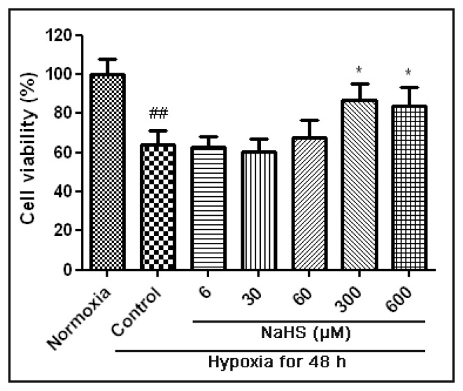
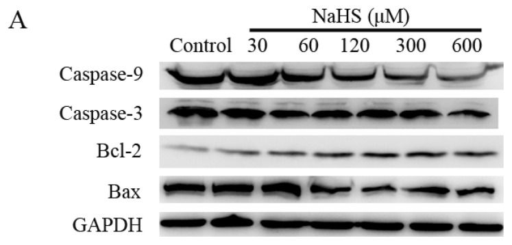
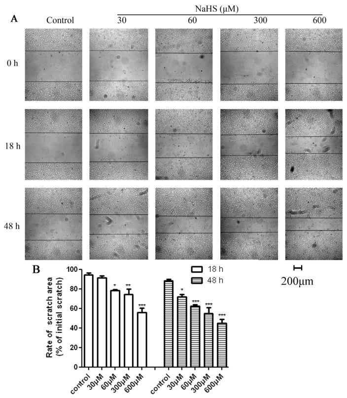
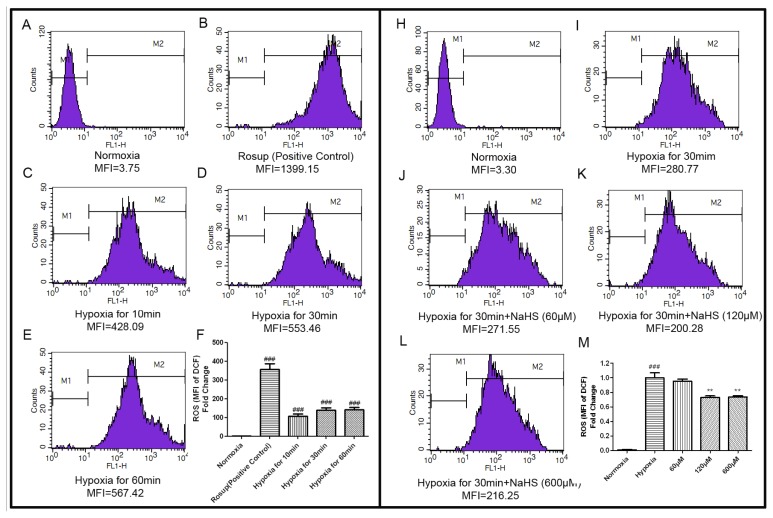
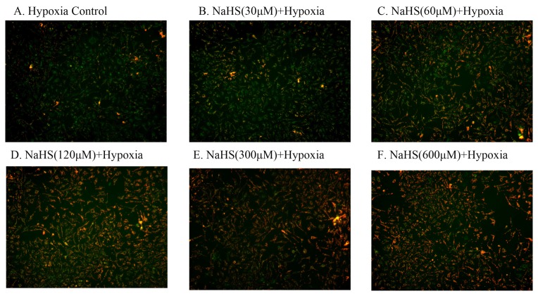
The aim of the study was to investigate the protective effects of sodium hydrosulfide (NaHS), a H2S donor, against hypoxia-induced injury in human umbilical vein endothelial cells (HUVECs) and also to look into the possible mechanisms by which H2S exerts this protective effect. 3-(4,5-dimethylthiazol-2-yl)-2,5-diphenyltetrazolium bromide (MTT) assay and scratch wound healing assay were chosen to measure the cell viability and migration-promoting effects. The fluorescent probe, DCFH-DA and 5,5',6,6'-Tetrachloro-1,1',3,3'-tetraethyl-imidacarbocyanine iodide (JC-1) were applied to detect the reactive oxygen species (ROS) level and mitochondrial membrane potential (ΔΨm). Furthermore, western blots were used to measure the expressions of the apoptosis-related proteins. Under hypoxic conditions, 300 μM and 600 μM of H2S could protect HUVECs against hypoxia-induced injury, as determined by MTT assay. Following the treatment of 60 µM NaHS for 18 h, scratch wound healing assays indicated that the scratch became much narrower than control group. After treatment with 60 µM, 120 µM, and 600 µM NaHS, and hypoxia for 30 min, flow cytometry demonstrated that the ROS concentrations decreased to 95.08% ± 5.52%, 73.14% ± 3.36%, and 73.51% ± 3.05%, respectively, compared with the control group. In addition, the JC-1 assay showed NaHS had a protective effect on mitochondria damage. Additionally, NaHS increased Bcl-2 expression and decreased the expression of Bax, Caspase-3 and Caspase-9 in a dose-dependent way. Our results suggest that H2S can protect endothelial cells and promote migration under hypoxic condition in HUVECs. These effects are partially associated with the preservation of mitochondrial function mediated by regulating the mitochondrial-dependent apoptotic pathway.
本研究旨在探讨硫化氢供体硫氢化钠(NaHS)对人脐静脉内皮细胞(HUVECs)缺氧诱导损伤的保护作用,并探究硫化氢发挥这种保护作用的可能机制。选用3-(4,5-二甲基噻唑-2-基)-2,5-二苯基四氮唑溴盐(MTT)法和划痕愈合试验来检测细胞活力和促迁移作用。应用荧光探针2',7'-二氯二氢荧光素二乙酸酯(DCFH-DA)和5,5',6,6'-四氯-1,1',3,3'-四乙基苯并咪唑羰花青碘化物(JC-1)检测活性氧(ROS)水平和线粒体膜电位(ΔΨm)。此外,采用蛋白质免疫印迹法检测凋亡相关蛋白的表达。MTT法检测结果显示,在缺氧条件下,300μM和600μM的硫化氢可保护HUVECs免受缺氧诱导的损伤。用60μM NaHS处理18小时后,划痕愈合试验表明划痕比对照组明显变窄。用60μM、120μM和600μM NaHS处理并缺氧30分钟后,流式细胞术显示,与对照组相比,ROS浓度分别降至95.08%±5.52%、73.14%±3.36%和73.51%±3.05%。此外,JC-1试验表明NaHS对线粒体损伤具有保护作用。另外,NaHS以剂量依赖的方式增加Bcl-2表达,降低Bax、Caspase-3和Caspase-9的表达。我们的结果表明,硫化氢可在缺氧条件下保护内皮细胞并促进其迁移。这些作用部分与通过调节线粒体依赖性凋亡途径介导的线粒体功能的维持有关。