Rusu Ligia D, Cosma Germina G H, Cernaianu Sorina M, Marin Mihnea N, Rusu Petre Florinel A, Ciocănescu Daniel P, Neferu Florin N
J Neuroeng Rehabil. 2013 Jul 3;10:67. doi: 10.1186/1743-0003-10-67.
Within the structure of the skeletal muscle, there are fascicles of muscular fibers that are made up of serially distributed contractile elements. These elements are controlled by the nervous system, control which results in obtaining the muscular strength required for movement and its control. This study presents the neuromuscular assessment using tensiomyography method (TMG).
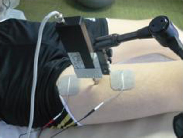
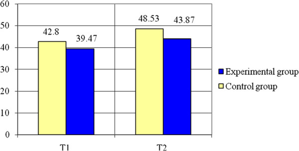
We studied two groups of soccer junior players, group 1 (experimental group) and group 2 (control group), each containing 15 soccer players; we have considered two situations of muscle training: the combination between the isometric-concentric contraction for group 1 and the concentric contraction for group 2. TMG is the electrical stimulation of the muscle group and the recording of the muscle parameters resulting after the isometric contraction: time contraction (Tc) and displacement (Dm) at rectus femoris muscle (RF), pointing out two moments T1 and T2.
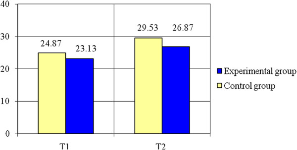
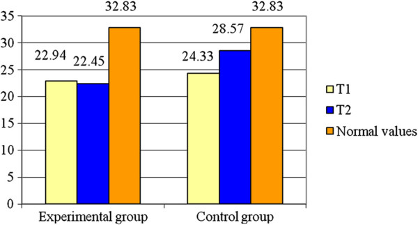
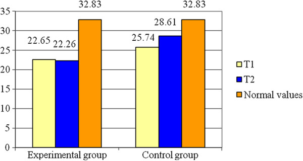
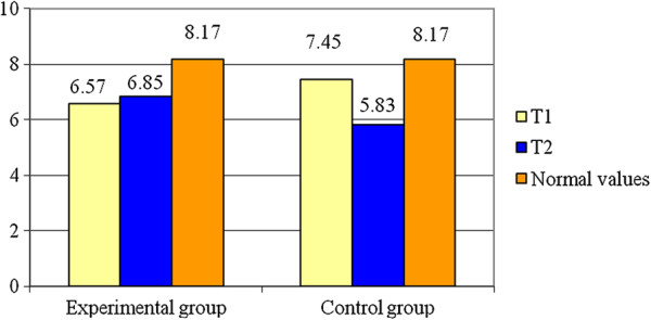
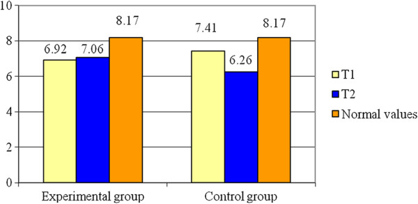
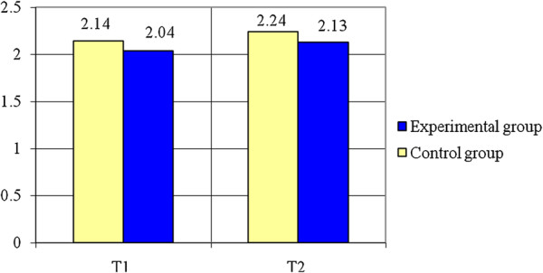
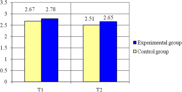
Tc decreasing and the Dm increasing involve a good response following the muscle training. For group 1, the Tc evolution is 22.54 ms/22.45 ms (T1/T2) for the right RF and 22.65 ms/22.26 ms for the left RF, while for group 2 results in a Tc evolution of 24.33 ms/28.57 ms (T1/T2) for the right RF and 25.74 ms/28.61 ms for the left RF. Dm for group 1, results in a 6.57 mm/6.85 mm (T1/T2) for the right RF and 6.92 mm/7.06 mm for the left RF, while for group 2, the Dm evolution shows 7.45 mm/5.83 mm (T1/T) for the right RF and 7.41 mm/6.26 mm for the left RF. Also, the evaluation on motor test indicated better results on T2 for the experimental group. Summarizing the results of Student t-test, we found significant differences between the averages of the two groups in all parameters (p < 0.001), the experimental group registering better results than the control one.
It is possible to develop muscle training which can be monitored through TMG.
在骨骼肌结构中,存在由一系列分布的收缩元件组成的肌纤维束。这些元件受神经系统控制,这种控制使得能够获得运动所需的肌肉力量及其控制。本研究介绍了使用张力肌电图法(TMG)进行的神经肌肉评估。
我们研究了两组青少年足球运动员,第1组(实验组)和第2组(对照组),每组各有15名足球运动员;我们考虑了两种肌肉训练情况:第1组为等长 - 向心收缩组合,第2组为向心收缩。TMG是对肌肉群进行电刺激,并记录等长收缩后产生的肌肉参数:股直肌(RF)的收缩时间(Tc)和位移(Dm),指出两个时刻T1和T2。
Tc降低和Dm增加表明肌肉训练后有良好反应。对于第1组,右侧RF的Tc变化为22.54毫秒/22.45毫秒(T1/T2),左侧RF为22.65毫秒/22.26毫秒,而对于第2组,右侧RF的Tc变化为24.33毫秒/28.57毫秒(T1/T2),左侧RF为25.74毫秒/28.61毫秒。第1组右侧RF的Dm为6.57毫米/6.85毫米(T1/T2),左侧RF为6.92毫米/7.06毫米,而对于第2组,右侧RF的Dm变化为7.45毫米/5.83毫米(T1/T),左侧RF为7.41毫米/6.26毫米。此外,运动测试评估表明实验组在T2时结果更好。总结学生t检验结果,我们发现两组在所有参数的平均值上存在显著差异(p < 0.001),实验组的结果优于对照组。
开发可通过TMG监测的肌肉训练是可能的。