Department of Radiation Oncology, Ajou University School of Medicine, Suwon, Republic of Korea.
Radiat Oncol. 2013 Jul 3;8:163. doi: 10.1186/1748-717X-8-163.
Localization of the tumor bed of breast cancer is crucial for accurate planning of boost irradiation. Lumpectomy cavity and surgical clips provide localizing information about tumor bed. However, defining the tumor bed is often difficult because of presence of unclear lumpectomy cavity and lack of certain information such as absence of surgical clips. In the present study, we evaluated the feasibility of initial diagnostic PET-CT in localization of the tumor bed using deformable image registration (DIR).
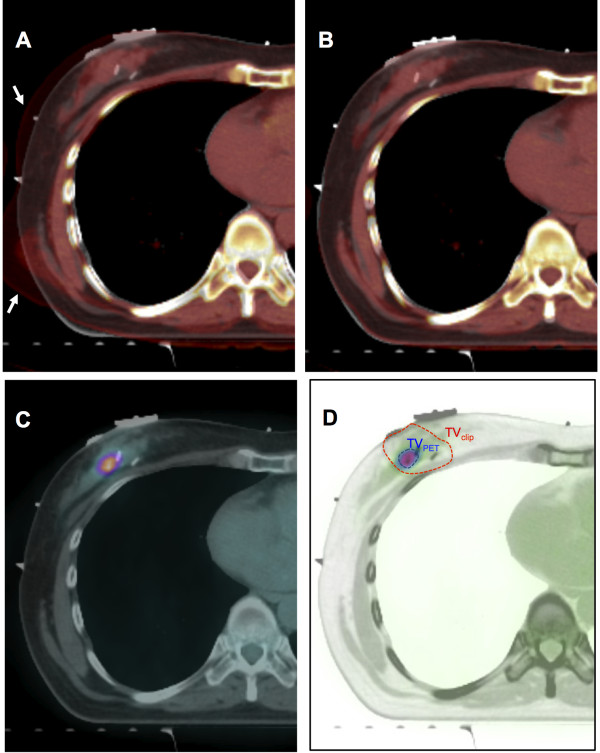
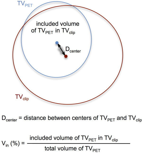
We selected twenty-five patients who had an initial diagnostic PET-CT performed and underwent breast-conserving surgery with surgical clips in tumor bed. In every individual patient, two target volumes were separately delineated on planning CT; 1) target volume based on surgical clips with a margin of 1 cm (TV(clip)) and 2) tumor volume based on 90% of maximum SUV on PET-CT registered by DIR (TV(PET)). The percent of TV(PET) in TV(clip) (V(in)) was calculated and distance between center points of two volumes (D(center)) was also measured.
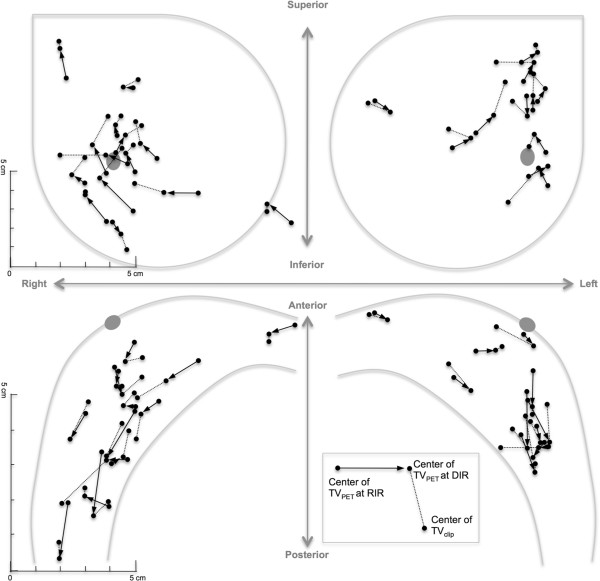
Mean D(center) between two volumes was 1.4 cm (range, 0.33-2.53). Mean V(in) was 94.8% (range, 60.9-100) and 100% in 18 out of 25 patients. When compared to the center of TV(clip), the center of TV(PET) tended to be located posteriorly (mean 0.3 cm, standard deviation 0.6), laterally (mean 0.3 cm, standard deviation 0.8) and inferiorly (mean 0.4 cm, standard deviation 0.9).
Initial diagnostic PET-CT can be one of the possible references to localize the tumor bed in breast cancer radiotherapy.
乳腺癌肿瘤床的定位对于准确规划加量照射至关重要。乳房切除术腔和手术夹提供了肿瘤床的定位信息。然而,由于存在不明确的乳房切除术腔和缺乏某些信息(如没有手术夹),定义肿瘤床通常很困难。在本研究中,我们评估了使用可变形图像配准(DIR)对初始诊断 PET-CT 定位肿瘤床的可行性。
我们选择了 25 名接受初始诊断 PET-CT 检查并在肿瘤床中接受乳腺保留手术和手术夹的患者。在每个患者中,分别在计划 CT 上勾画两个靶区;1)基于手术夹的靶区,边界为 1cm(TV(clip))和 2)基于 90%最大 SUV 的肿瘤体积在 PET-CT 上由 DIR 注册(TV(PET))。计算 TV(PET)在 TV(clip)中的百分比(V(in)),并测量两个体积中心点之间的距离(D(center))。
两个体积之间的平均 D(center)为 1.4cm(范围,0.33-2.53)。平均 V(in)为 94.8%(范围,60.9-100),25 名患者中有 18 名达到 100%。与 TV(clip)的中心点相比,TV(PET)的中心点倾向于位于后部(平均 0.3cm,标准差 0.6)、侧面(平均 0.3cm,标准差 0.8)和下方(平均 0.4cm,标准差 0.9)。
初始诊断 PET-CT 可以成为乳腺癌放射治疗中定位肿瘤床的一种可能参考。