• 文献检索
  • 文档翻译
  • 深度研究
  • 学术资讯
  • Suppr Zotero 插件Zotero 插件
  • 邀请有礼
  • 套餐&价格
  • 历史记录
应用&插件
Suppr Zotero 插件Zotero 插件浏览器插件Mac 客户端Windows 客户端微信小程序
定价
高级版会员购买积分包购买API积分包
服务
文献检索文档翻译深度研究API 文档MCP 服务
关于我们
关于 Suppr公司介绍联系我们用户协议隐私条款
关注我们

Suppr 超能文献

核心技术专利:CN118964589B侵权必究
粤ICP备2023148730 号-1Suppr @ 2026

文献检索

告别复杂PubMed语法,用中文像聊天一样搜索,搜遍4000万医学文献。AI智能推荐,让科研检索更轻松。

立即免费搜索

文件翻译

保留排版,准确专业,支持PDF/Word/PPT等文件格式,支持 12+语言互译。

免费翻译文档

深度研究

AI帮你快速写综述,25分钟生成高质量综述,智能提取关键信息,辅助科研写作。

立即免费体验

弥散张量纤维束成像与容积成像在行为变异型额颞叶痴呆诊断中的比较。

Diffusion tensor tractography versus volumetric imaging in the diagnosis of behavioral variant frontotemporal dementia.

机构信息

Clinical Memory Research Unit, Department of Clinical Medicine, Lund University, Lund, Sweden.

出版信息

PLoS One. 2013 Jul 18;8(7):e66932. doi: 10.1371/journal.pone.0066932. Print 2013.

DOI:10.1371/journal.pone.0066932
PMID:23874403
原文链接:https://pmc.ncbi.nlm.nih.gov/articles/PMC3715470/
Abstract

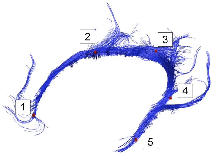
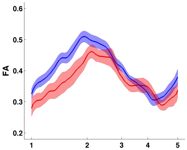
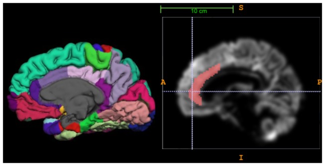
MRI diffusion tensor imaging (DTI) studies of white matter integrity in behavioral variant frontotemporal dementia have consistently shown involvement of frontal and temporal white matter, corresponding to regional loss of cortical volume. Volumetric imaging has a suboptimal sensitivity as a diagnostic tool and thus we wanted to explore if DTI is a better method to discriminate patients and controls than volumetric imaging. We examined the anterior cingulum bundle in 14 patients with behavioral variant frontotemporal dementia and 22 healthy controls using deterministic manual diffusion tensor tractography, and compared DTI parameters with two measures of cortical atrophy, VBM and cortical thickness, of the anterior cingulate cortex (ACC). Statistically significant changes between patients and controls were detected in all DTI parameters, with large effect sizes. ROC-AUC was for the best DTI parameters: 0.92 (fractional anisotropy) to 0.97 (radial diffusivity), 0.82 for the best cortical parameter, VBM of the ACC. Results from the AUC were confirmed with binary logistic regression analysis including demographic variables, but only for fractional anisotropy and mean diffusivity. Ability to classify patient/nonpatient status was significantly better for mean diffusivity vs. VBM (p=0.031), and borderline significant for fractional anisotropy vs. VBM (p=0.062). The results indicate that DTI could offer advantages in comparison with the assessment of cortical volume in differentiating patients with behavioral variant frontotemporal dementia and controls.

摘要

MRI 弥散张量成像(DTI)研究行为变异型额颞叶痴呆的脑白质完整性,一致显示额叶和颞叶白质受累,与皮质体积的区域性丧失相对应。容积成像作为一种诊断工具的灵敏度较低,因此我们想探讨 DTI 是否比容积成像更能区分患者和对照者。我们使用确定性手动弥散张量跟踪法检查了 14 例行为变异型额颞叶痴呆患者和 22 名健康对照者的前扣带束,将 DTI 参数与前扣带皮质(ACC)的皮质体积测量(VBM)和皮质厚度两种皮质萎缩测量方法进行了比较。在所有 DTI 参数中,患者与对照组之间均检测到统计学上显著的变化,具有较大的效应量。最佳 DTI 参数的 ROC-AUC 为:0.92(各向异性分数)至 0.97(辐射状弥散系数),最佳皮质参数为 0.82(ACC 的 VBM)。AUC 的结果得到了包括人口统计学变量的二元逻辑回归分析的证实,但仅针对各向异性分数和平均弥散系数。与 VBM 相比,平均弥散系数对区分患者/非患者状态的能力明显更好(p=0.031),与 VBM 相比,各向异性分数的结果接近显著(p=0.062)。结果表明,与评估皮质体积相比,DTI 在区分行为变异型额颞叶痴呆患者和对照者方面可能具有优势。

https://cdn.ncbi.nlm.nih.gov/pmc/blobs/bf30/3715470/8115e89aeb0b/pone.0066932.g003.jpg
https://cdn.ncbi.nlm.nih.gov/pmc/blobs/bf30/3715470/dc2c6b703205/pone.0066932.g001.jpg
https://cdn.ncbi.nlm.nih.gov/pmc/blobs/bf30/3715470/e173388f8dd4/pone.0066932.g002.jpg
https://cdn.ncbi.nlm.nih.gov/pmc/blobs/bf30/3715470/8115e89aeb0b/pone.0066932.g003.jpg
https://cdn.ncbi.nlm.nih.gov/pmc/blobs/bf30/3715470/dc2c6b703205/pone.0066932.g001.jpg
https://cdn.ncbi.nlm.nih.gov/pmc/blobs/bf30/3715470/e173388f8dd4/pone.0066932.g002.jpg
https://cdn.ncbi.nlm.nih.gov/pmc/blobs/bf30/3715470/8115e89aeb0b/pone.0066932.g003.jpg

相似文献

1
Diffusion tensor tractography versus volumetric imaging in the diagnosis of behavioral variant frontotemporal dementia.弥散张量纤维束成像与容积成像在行为变异型额颞叶痴呆诊断中的比较。
PLoS One. 2013 Jul 18;8(7):e66932. doi: 10.1371/journal.pone.0066932. Print 2013.
2
An advanced white matter tract analysis in frontotemporal dementia and early-onset Alzheimer's disease.额颞叶痴呆和早发性阿尔茨海默病的高级白质束分析
Brain Imaging Behav. 2016 Dec;10(4):1038-1053. doi: 10.1007/s11682-015-9458-5.
3
Hemispheric Regional Based Analysis of Diffusion Tensor Imaging and Diffusion Tensor Tractography in Patients with Temporal Lobe Epilepsy and Correlation with Patient outcomes.基于半球区域的分析弥散张量成像和弥散张量纤维束成像在颞叶癫痫患者中的应用及其与患者预后的相关性。
Sci Rep. 2019 Jan 18;9(1):215. doi: 10.1038/s41598-018-36818-x.
4
Diffusion parameters of the core of cingulum are associated with age-related ventricular enlargement: a diffusion tensor tractography study.扣带束核心的扩散参数与年龄相关的心室扩大有关:一项扩散张量纤维束成像研究。
Neuroradiology. 2018 Oct;60(10):1013-1018. doi: 10.1007/s00234-018-2068-3. Epub 2018 Aug 8.
5
A combined diffusion tensor imaging and magnetic resonance spectroscopy study of patients with schizophrenia.一项针对精神分裂症患者的弥散张量成像与磁共振波谱联合研究。
Schizophr Res. 2016 Feb;170(2-3):341-50. doi: 10.1016/j.schres.2015.12.003. Epub 2015 Dec 21.
6
Diagnostic Value of Diffusion Tensor Imaging and Positron Emission Tomography in Early Stages of Frontotemporal Dementia.弥散张量成像和正电子发射断层扫描在额颞叶痴呆早期的诊断价值。
J Alzheimers Dis. 2018;63(1):239-253. doi: 10.3233/JAD-170224.
7
Gray and white matter water diffusion in the syndromic variants of frontotemporal dementia.额颞叶痴呆综合征变异型的灰白质水弥散。
Neurology. 2010 Apr 20;74(16):1279-87. doi: 10.1212/WNL.0b013e3181d9edde.
8
Longitudinal diffusion tensor imaging in frontotemporal dementia.额颞叶痴呆的纵向扩散张量成像。
Ann Neurol. 2015 Jan;77(1):33-46. doi: 10.1002/ana.24296. Epub 2014 Nov 17.
9
Regional structural hypo- and hyperconnectivity of frontal-striatal and frontal-thalamic pathways in behavioral variant frontotemporal dementia.行为变异型额颞叶痴呆患者额纹状体和额丘脑通路上的区域性结构连接低下和连接过度。
Hum Brain Mapp. 2018 Oct;39(10):4083-4093. doi: 10.1002/hbm.24233. Epub 2018 Jun 20.
10
Partial volume effect as a hidden covariate in DTI analyses.部分容积效应作为 DTI 分析中的隐藏协变量。
Neuroimage. 2011 Apr 15;55(4):1566-76. doi: 10.1016/j.neuroimage.2011.01.048. Epub 2011 Jan 22.

引用本文的文献

1
Multiparametric MRI-based biomarkers in the non-fluent and semantic variants of primary progressive aphasia.基于多参数磁共振成像的原发性进行性失语非流利型和语义型变体生物标志物
J Neurol. 2025 Jul 2;272(8):490. doi: 10.1007/s00415-025-13215-9.
2
Multiclass characterization of frontotemporal dementia variants via multimodal brain network computational inference.通过多模态脑网络计算推理对额颞叶痴呆变体进行多类别特征描述。
Netw Neurosci. 2023 Jan 1;7(1):322-350. doi: 10.1162/netn_a_00285. eCollection 2023.
3
Alterations of cerebellar white matter integrity and associations with cognitive impairments in schizophrenia.

本文引用的文献

1
Spatial analysis of diffusion tensor tractography statistics along the inferior fronto-occipital fasciculus with application in progressive supranuclear palsy.基于弥散张量纤维束追踪技术的下额枕束空间分析及其在进行性核上性麻痹中的应用。
MAGMA. 2013 Dec;26(6):527-37. doi: 10.1007/s10334-013-0368-5. Epub 2013 Mar 30.
2
MRI signatures of brain macrostructural atrophy and microstructural degradation in frontotemporal lobar degeneration subtypes.MRI 特征显示额颞叶变性各亚型的脑宏观结构萎缩和微观结构退化。
J Alzheimers Dis. 2013;33(2):431-44. doi: 10.3233/JAD-2012-121156.
3
Gray matter atrophy in behavioral variant frontotemporal dementia: a meta-analysis of voxel-based morphometry studies.
精神分裂症中小脑白质完整性的改变及其与认知障碍的关联。
Front Psychiatry. 2022 Sep 26;13:993866. doi: 10.3389/fpsyt.2022.993866. eCollection 2022.
4
A multicentre validation study of the diagnostic value of plasma neurofilament light.血浆神经丝轻链的诊断价值的多中心验证研究。
Nat Commun. 2021 Jun 7;12(1):3400. doi: 10.1038/s41467-021-23620-z.
5
Neuroimaging in Frontotemporal Lobar Degeneration: Research and Clinical Utility.额颞叶变性的神经影像学:研究与临床应用。
Adv Exp Med Biol. 2021;1281:93-112. doi: 10.1007/978-3-030-51140-1_7.
6
Plasma neurofilament light protein correlates with diffusion tensor imaging metrics in frontotemporal dementia.血浆神经丝轻蛋白与额颞叶痴呆的弥散张量成像指标相关。
PLoS One. 2020 Oct 27;15(10):e0236384. doi: 10.1371/journal.pone.0236384. eCollection 2020.
7
Relationship between Visual Perception and Microstructural Change of the Superior Longitudinal Fasciculus in Patients with Brain Injury in the Right Hemisphere: A Preliminary Diffusion Tensor Tractography Study.右侧半球脑损伤患者视觉感知与上纵束微结构变化的关系:一项初步的扩散张量纤维束成像研究
Diagnostics (Basel). 2020 Aug 27;10(9):641. doi: 10.3390/diagnostics10090641.
8
Structural Characteristic of the Arcuate Fasciculus in Patients with Fluent Aphasia Following Intracranial Hemorrhage: A Diffusion Tensor Tractography Study.颅内出血后流利性失语患者弓状束的结构特征:一项扩散张量纤维束成像研究
Brain Sci. 2020 May 6;10(5):280. doi: 10.3390/brainsci10050280.
9
Injury of Corticospinal Tract in a Patient with Subarachnoid Hemorrhage as Determined by Diffusion Tensor Tractography: A Case Report.基于扩散张量纤维束成像确定的蛛网膜下腔出血患者皮质脊髓束损伤:一例报告
Brain Sci. 2020 Mar 19;10(3):177. doi: 10.3390/brainsci10030177.
10
Diffusion-Tensor-Tractography-Based Diagnosis for Injury of Corticospinal Tract in a Patient with Hemiplegia Following Traumatic Brain Injury.基于扩散张量纤维束成像的创伤性脑损伤后偏瘫患者皮质脊髓束损伤诊断
Diagnostics (Basel). 2020 Mar 13;10(3):156. doi: 10.3390/diagnostics10030156.
行为变异型额颞叶痴呆的灰质萎缩:基于体素形态计量学研究的荟萃分析。
Dement Geriatr Cogn Disord. 2012;33(2-3):141-8. doi: 10.1159/000338176. Epub 2012 May 7.
4
A comparison between volumetric data generated by voxel-based morphometry and manual parcellation of multimodal regions of the frontal lobe.基于体素的形态计量学生成的容积数据与额叶多模态区域的手动分割的比较。
AJNR Am J Neuroradiol. 2012 Nov;33(10):1957-63. doi: 10.3174/ajnr.A3093. Epub 2012 May 10.
5
Neuroimaging signatures of frontotemporal dementia genetics: C9ORF72, tau, progranulin and sporadics.额颞叶痴呆遗传学的神经影像学特征:C9ORF72、tau、颗粒蛋白和散发性病例。
Brain. 2012 Mar;135(Pt 3):794-806. doi: 10.1093/brain/aws001.
6
White matter damage in frontotemporal lobar degeneration spectrum.额颞叶变性谱中的脑白质损伤。
Cereb Cortex. 2012 Dec;22(12):2705-14. doi: 10.1093/cercor/bhr288. Epub 2011 Oct 10.
7
Sensitivity of revised diagnostic criteria for the behavioural variant of frontotemporal dementia.修订后的额颞叶痴呆行为变异型诊断标准的敏感性。
Brain. 2011 Sep;134(Pt 9):2456-77. doi: 10.1093/brain/awr179. Epub 2011 Aug 2.
8
Convergent grey and white matter evidence of orbitofrontal cortex changes related to disinhibition in behavioural variant frontotemporal dementia.与行为变异额颞叶痴呆相关的眶额皮质去抑制改变的会聚灰质和白质证据。
Brain. 2011 Sep;134(Pt 9):2502-12. doi: 10.1093/brain/awr173. Epub 2011 Jul 23.
9
Joint assessment of structural, perfusion, and diffusion MRI in Alzheimer's disease and frontotemporal dementia.阿尔茨海默病和额颞叶痴呆中结构、灌注和扩散磁共振成像的联合评估
Int J Alzheimers Dis. 2011;2011:546871. doi: 10.4061/2011/546871. Epub 2011 Jun 27.
10
Partial volume effect as a hidden covariate in DTI analyses.部分容积效应作为 DTI 分析中的隐藏协变量。
Neuroimage. 2011 Apr 15;55(4):1566-76. doi: 10.1016/j.neuroimage.2011.01.048. Epub 2011 Jan 22.