Liver Int. 2013 Nov;33(10):1566-74. doi: 10.1111/liv.12238.
BACKGROUND & AIMS: Apoptosis mediated by p53 plays a pathological role in the progression of hepatosteatosis. It is noteworthy that p53 can promote the expression of damage-regulated autophagy modulator (DRAM), an inducer of autophagy-mediated apoptosis. However, the relationship between p53-mediated apoptosis and autophagy in hepatosteatosis remains elusive. This study aimed to examine how p53 orchestrates autophagy and apoptosis to affect hepatosteatosis.
HepG2 cells were treated with oleic acid (OA) for 24 h to induce hepatosteatosis. Mice were fed a high-fat diet for 20 or 40 weeks to induce hepatosteatosis.
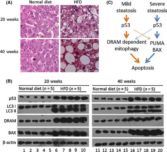
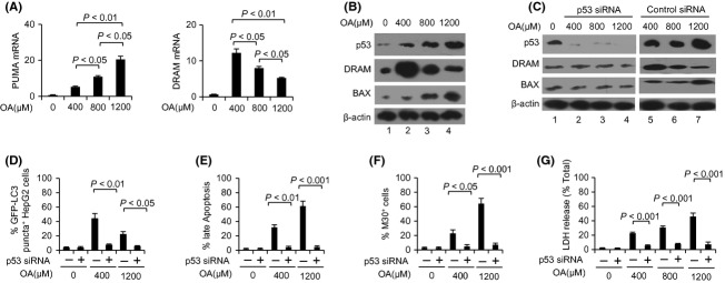
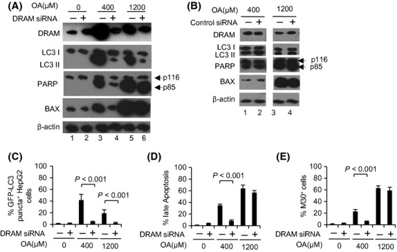
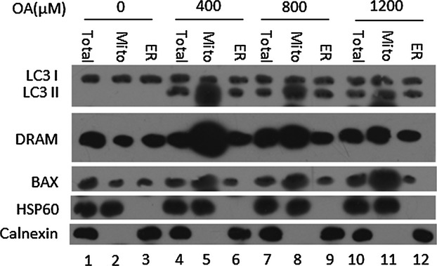
OA induced a dose-dependent increase in steatosis severity and apoptosis. OA also induced autophagy, which was a critical inducer of apoptosis in mild steatosis induced by 400 μM OA, but not in the more severe steatosis induced by 800 and 1200 μM OA. p53 inhibition by siRNA mostly blocked OA-induced apoptosis and autophagy. Moreover, OA-induced autophagy was DRAM-dependent and primarily occurred in the mitochondria (mitophagy), where DRAM was localized. In severe steatosis induced by 1200 μM OA, apoptosis was mainly dependent on p53-induced expression of BAX, which was also localized to the mitochondria. Our in vivo study showed that p53 expression increased in both mild and severe hepatosteatosis. Increased DRAM expression and autophagy were identified in mild hepatosteatosis, whereas greater BAX expression was observed in severe hepatosteatosis.
p53 may induce apoptosis via different mechanisms. DRAM-mediated mitophagy is a primary apoptotic inducer in mild hepatosteatosis, whereas p53-induced BAX expression mainly induces apoptosis in severe hepatosteatosis.
p53 介导的细胞凋亡在脂肪性肝炎的进展中起着病理性作用。值得注意的是,p53 可以促进损伤调节自噬调节剂(DRAM)的表达,DRAM 是自噬介导细胞凋亡的诱导剂。然而,p53 介导的细胞凋亡与脂肪性肝炎中的自噬之间的关系仍不清楚。本研究旨在探讨 p53 如何协调自噬和细胞凋亡来影响脂肪性肝炎。
用油酸(OA)处理 HepG2 细胞 24 小时以诱导脂肪性肝炎。用高脂肪饮食喂养小鼠 20 或 40 周以诱导脂肪性肝炎。
OA 诱导的脂肪变性严重程度和细胞凋亡呈剂量依赖性增加。OA 还诱导自噬,在 400 μM OA 诱导的轻度脂肪变性中,自噬是细胞凋亡的关键诱导剂,但在 800 和 1200 μM OA 诱导的更严重脂肪变性中则不是。siRNA 抑制 p53 可阻断 OA 诱导的细胞凋亡和自噬。此外,OA 诱导的自噬依赖于 DRAM,主要发生在线粒体(线粒体自噬)中,DRAM 定位于线粒体。在 1200 μM OA 诱导的严重脂肪变性中,细胞凋亡主要依赖于 p53 诱导的 BAX 表达,BAX 也定位于线粒体。我们的体内研究表明,p53 在轻度和重度脂肪性肝炎中表达增加。在轻度脂肪性肝炎中发现 DRAM 表达增加和自噬增加,而在重度脂肪性肝炎中则观察到更大的 BAX 表达。
p53 可能通过不同的机制诱导细胞凋亡。DRAM 介导的线粒体自噬是轻度脂肪性肝炎中主要的凋亡诱导剂,而 p53 诱导的 BAX 表达主要诱导重度脂肪性肝炎中的凋亡。