Biological Faculty, Sofia University "Saint Kliment Ohridski", 8 Dragan Tzankov str, Sofia 1164, Bulgaria.
Int J Mol Sci. 2013 Jul 22;14(7):15121-40. doi: 10.3390/ijms140715121.
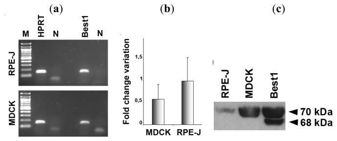
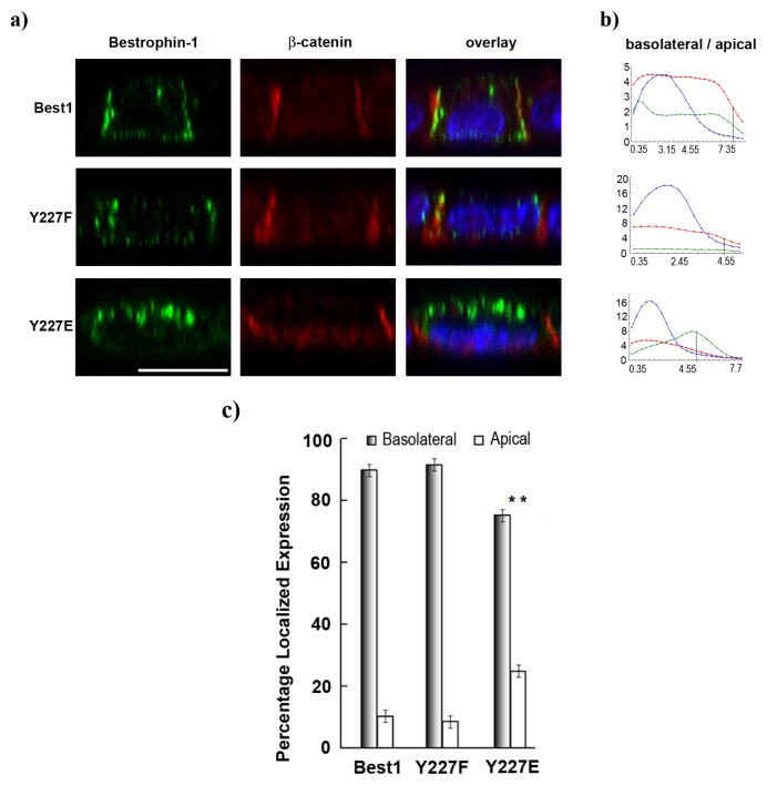
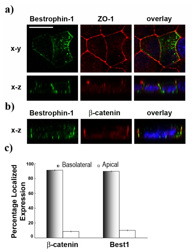
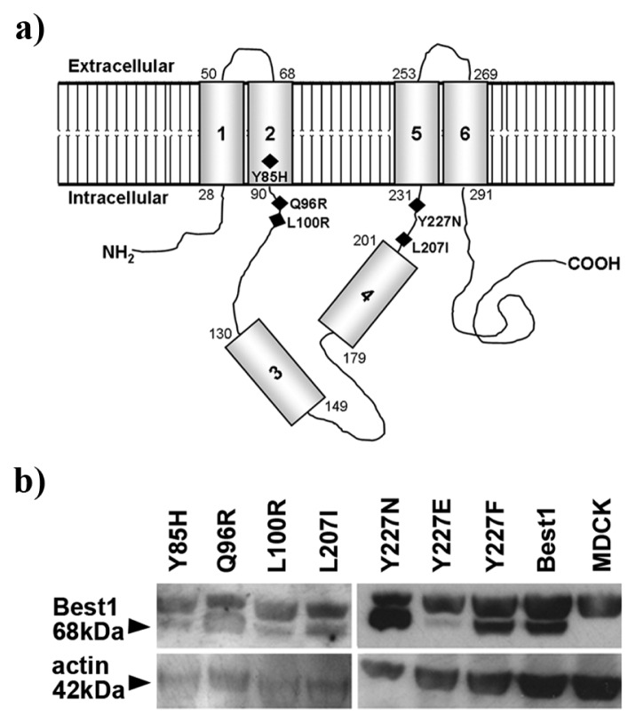
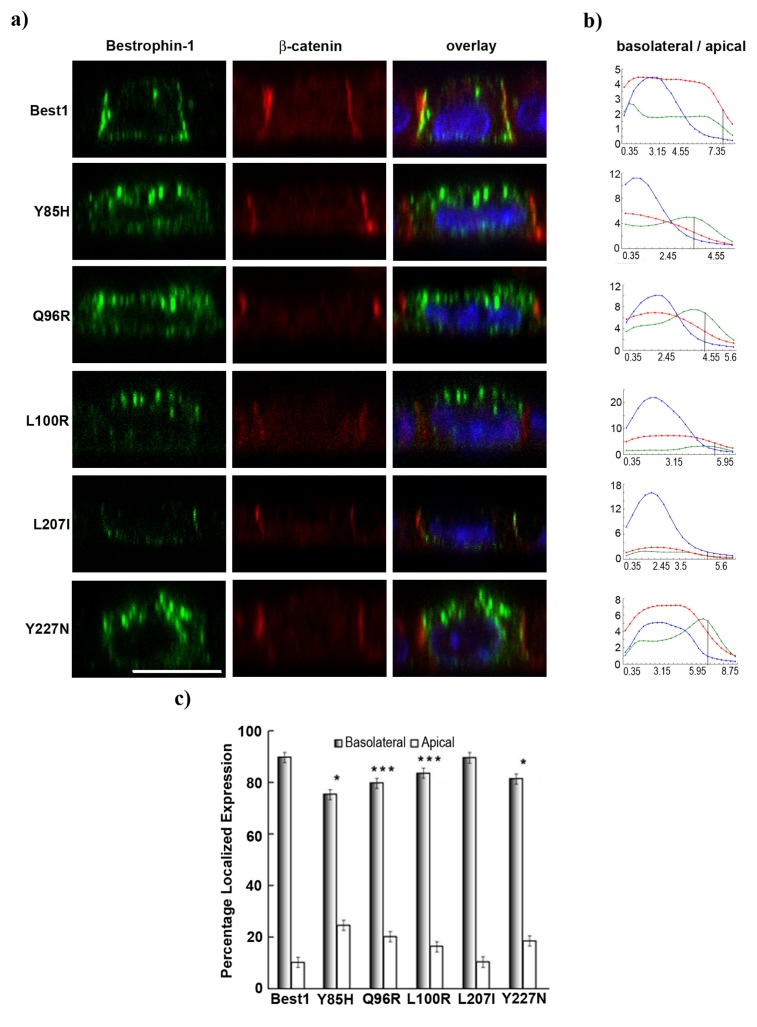
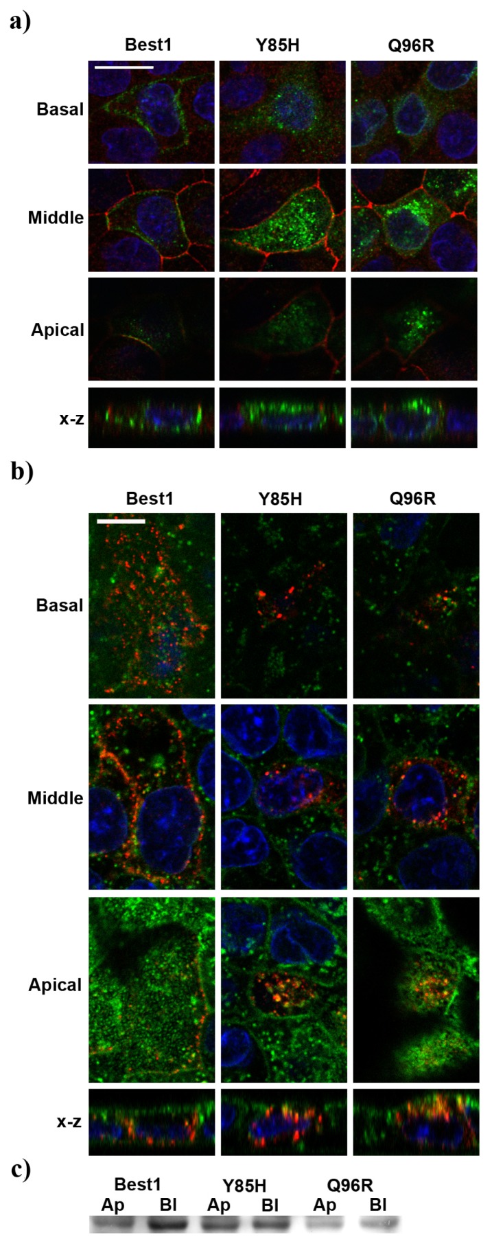
Mutations in BEST1 gene, encoding the bestrophin-1 (Best1) protein are associated with macular dystrophies. Best1 is predominantly expressed in the retinal pigment epithelium (RPE), and is inserted in its basolateral membrane. We investigated the cellular localization in polarized MDCKII cells of disease-associated Best1 mutant proteins to study specific sorting motifs of Best1. Real-time PCR and western blots for endogenous expression of BEST1 in MDCK cells were performed. Best1 mutant constructs were generated using site-directed mutagenesis and transfected in MDCK cells. For protein sorting, confocal microscopy studies, biotinylation assays and statistical methods for quantification of mislocalization were used. Analysis of endogenous expression of BEST1 in MDCK cells revealed the presence of BEST1 transcript but no protein. Confocal microscopy and quantitative analyses indicate that transfected normal human Best1 displays a basolateral localization in MDCK cells, while cell sorting of several Best1 mutants (Y85H, Q96R, L100R, Y227N, Y227E) was altered. In contrast to constitutively active Y227E, constitutively inactive Y227F Best1 mutant localized basolaterally similar to the normal Best1 protein. Our data suggest that at least three basolateral sorting motifs might be implicated in proper Best1 basolateral localization. In addition, non-phosphorylated tyrosine 227 could play a role for basolateral delivery.
BEST1 基因突变与黄斑营养不良有关,该基因编码的蛋白为 bestrophin-1(Best1)。Best1 主要在视网膜色素上皮(RPE)中表达,并插入其基底外侧膜。我们研究了与疾病相关的 Best1 突变蛋白在极化 MDCKII 细胞中的细胞定位,以研究 Best1 的特定分拣基序。在 MDCK 细胞中进行 BEST1 内源性表达的实时 PCR 和 Western blot。使用定点诱变生成 Best1 突变构建体,并在 MDCK 细胞中转染。对于蛋白质分拣,使用共聚焦显微镜研究、生物素化测定和用于错误定位定量的统计方法。在 MDCK 细胞中分析 BEST1 的内源性表达显示存在 BEST1 转录本但没有蛋白质。共聚焦显微镜和定量分析表明,转染的正常人 Best1 在 MDCK 细胞中显示出基底外侧定位,而几种 Best1 突变体(Y85H、Q96R、L100R、Y227N、Y227E)的细胞分拣发生改变。与组成性激活的 Y227E 相反,组成性失活的 Y227F Best1 突变体与正常的 Best1 蛋白一样定位于基底外侧。我们的数据表明,至少有三个基底外侧分拣基序可能与 Best1 基底外侧定位有关。此外,非磷酸化的酪氨酸 227 可能在基底外侧递送上发挥作用。