Department of Molecular Genetics, Cardiovascular Research Institute Maastricht (CARIM), Maastricht, The Netherlands.
PLoS One. 2013 Jul 24;8(7):e68811. doi: 10.1371/journal.pone.0068811. Print 2013.
Enhancement of collateral development in coronary or peripheral artery disease is a therapeutic target, but it has proven difficult to achieve. Macrophages are key players in collateral remodeling, yet the effect of different macrophage subsets on arteriogenesis has not been investigated.
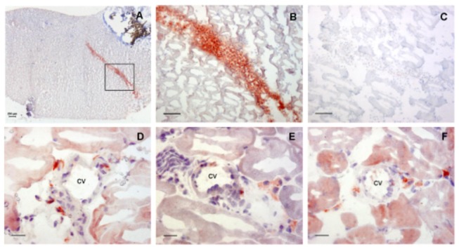
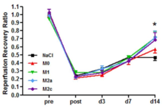
Murine macrophages were cultured from bone marrow and polarized into M1 (IFNγ), M2a (IL-4) or M2c (IL-10) subsets. C57BL/6 mice underwent femoral artery ligation followed by intramuscular injection of macrophage subsets. Using eGFP expressing macrophages, cells could be detected at least 6 days after ligation and were located in the perivascular space of collateral vessels. After 14 days, perfusion ratio was increased in animals treated with M1 as well as M2a and M2c macrophages compared to control. Depletion of circulating monocytes by clodronate liposome injections did not hamper reperfusion recovery, however, treatment with exogenous polarized macrophages improved perfusion ratio after 14 days again. We used IL10R(fl/fl)/LysMCre(+) mice to study the effect of inhibition of endogenous polarization towards specifically M2c macrophages on arteriogenesis. Deletion of the IL10-receptor (IL10R) in the myeloid lineage did not affect reperfusion recovery, yet the pro-arteriogenic effect of exogenously injected M2c macrophages was still present.
Local injection of polarized macrophages promotes reperfusion recovery after femoral artery ligation and is not influenced by depletion of circulatory monocytes. Preventing endogenous M2c polarization did not affect reperfusion recovery suggesting that M2c's are not required for collateralization, but are sufficient to induce collateral formation upon exogenous administration. This is the first study using local injection of macrophage subsets showing the pro-arteriogenic effect of polarized macrophages.
增强冠状动脉或外周动脉疾病的侧支血管发育是一种治疗靶点,但迄今为止难以实现。巨噬细胞是侧支血管重构的关键参与者,然而不同巨噬细胞亚群对动脉生成的影响尚未得到研究。
从骨髓中培养出小鼠巨噬细胞,并将其极化成为 M1(IFNγ)、M2a(IL-4)或 M2c(IL-10)亚群。C57BL/6 小鼠进行股动脉结扎,随后肌肉内注射巨噬细胞亚群。使用表达 eGFP 的巨噬细胞,在结扎后至少 6 天可以检测到细胞,并位于侧支血管的血管周围空间。14 天后,与对照组相比,用 M1 以及 M2a 和 M2c 巨噬细胞处理的动物的灌注比例增加。用氯膦酸盐脂质体注射耗竭循环单核细胞不会阻碍再灌注恢复,但再次用外源性极化巨噬细胞治疗 14 天后,灌注比例再次提高。我们使用 IL10R(fl/fl)/LysMCre(+) 小鼠来研究抑制内源性极化成特定的 M2c 巨噬细胞对动脉生成的影响。在髓系中缺失 IL10 受体(IL10R)不会影响再灌注恢复,但外源性注射的 M2c 巨噬细胞的促动脉生成作用仍然存在。
局部注射极化巨噬细胞可促进股动脉结扎后的再灌注恢复,且不受循环单核细胞耗竭的影响。阻止内源性 M2c 极化不会影响再灌注恢复,这表明 M2c 对侧支血管形成不是必需的,但在外源性给药时足以诱导侧支血管形成。这是首次使用局部注射巨噬细胞亚群研究极化巨噬细胞的促动脉生成作用的研究。