Mercer Robert R, Scabilloni James F, Hubbs Ann F, Battelli Lori A, McKinney Walter, Friend Sherri, Wolfarth Michael G, Andrew Michael, Castranova Vincent, Porter Dale W
Part Fibre Toxicol. 2013 Jul 30;10:33. doi: 10.1186/1743-8977-10-33.
Prior studies have demonstrated a rapid and progressive acute phase response to bolus aspiration of multi-walled carbon nanotubes (MWCNTs). In this study we sought to test the hypothesis that inhalation exposure to MWCNT produces a fibrotic response and that the response is chronically persistent. To address the hypothesis that inhaled MWCNTs cause persistent morphologic changes, male C57BL/6 J mice were exposed in a whole-body inhalation system to a MWCNT aerosol and the fibrotic response in the alveolar region examined at up to 336 days after termination of exposure.
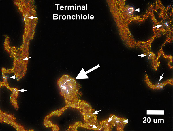
Inhalation exposure was to a 5 mg/m3 MWCNT aerosol for 5 hours/day for 12 days (4 times/week for 3 weeks). At the end of inhalation exposures, lungs were either lavaged for analysis of bronchoalveolar lavage (BAL) or preserved by vascular perfusion of fixative while inflated with air at 1, 14, 84, 168 and 336 days post inhalation exposure. Separate, clean-air control groups were also studied. Light microscopy, enhanced darkfield microscopy and field emission electron microscopy (FESEM) of tissue sections were used to analyze the distribution of lung burden following inhalation exposure. Morphometric measurements of Sirius Red staining for fibrillar collagen were used to assess the connective tissue response. Serial section analysis of enhanced darkfield microscope images was used to examine the redistribution of MWCNT fibers within the lungs during the post-exposure period.
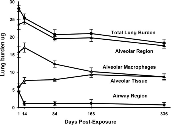
At day 1 post-exposure 84 ± 3 and 16 ± 2 percent of the lung burden (Mean ± S.E., N = 5) were in the alveolar and airway regions, respectively. Initial distribution within the alveolar region was 56 ± 5, 7 ± 4 and 20 ± 3 percent of lung burden in alveolar macrophages, alveolar airspaces and alveolar tissue, respectively. Clearance reduced the alveolar macrophage burden of MWCNTs by 35 percent between 1 and 168 days post-exposure, while the content of MWCNTs in the alveolar tissue increased by 63 percent. Large MWCNT structures containing greater than 4 fibers were 53.6 percent of the initial lung burden and accounted for the majority of the decline with clearance, while lung burden of singlet MWCNT was essentially unchanged. The mean linear intercept of alveolar airspace, a measure of the expansion of the lungs, was not significantly different between groups. Pulmonary inflammation and damage, measured as the number of polymorphnuclear leukocytes (PMNs) or lactate dehydrogenase activity (LDH) and albumin in BAL, increased rapidly (1 day post) after inhalation of MWCNTs and declined slowly with time post-exposure. The fibrillar collagen in the alveolar region of MWCNT-exposed mice demonstrated a progressive increase in thickness over time (0.17 ± 0.02, 0.22 ± 0.02, 0.26 ± 0.03, 0.25 ± 0.02 and 0.29 ± 0.01 microns for 1, 14, 84, 168 and 336 days post-exposure) and was significantly different from clean-air controls (0.16 ± 0.02) at 84 and (0.15 ± 0.02) at 336 days post-exposure.
Despite the relatively low fraction of the lung burden being delivered to the alveolar tissue, the average thickness of connective tissue in the alveolar region increased by 70% in the 336 days after inhalation exposure. These results demonstrate that inhaled MWCNTs deposit and are retained within the alveolar tissue where they produce a progressive and persistent fibrotic response up to 336 days post-exposure.

先前的研究已证明,多壁碳纳米管(MWCNT)团注吸入会引发快速且渐进的急性期反应。在本研究中,我们试图验证以下假设:吸入MWCNT会产生纤维化反应,且该反应具有慢性持续性。为验证吸入MWCNT会导致持续性形态学改变这一假设,将雄性C57BL/6 J小鼠置于全身吸入系统中,使其暴露于MWCNT气溶胶,并在暴露终止后长达336天的时间内检查肺泡区域的纤维化反应。
吸入暴露为5 mg/m3的MWCNT气溶胶,每天暴露5小时,持续12天(每周4次,共3周)。在吸入暴露结束时,对肺部进行灌洗以分析支气管肺泡灌洗(BAL),或者在吸入暴露后1、14、84、168和336天,通过向充气的肺中灌注固定剂来保存肺部。还研究了单独的清洁空气对照组。使用组织切片的光学显微镜、增强暗场显微镜和场发射电子显微镜(FESEM)分析吸入暴露后肺部负荷的分布。用Sirius Red染色对纤维状胶原进行形态计量学测量,以评估结缔组织反应。使用增强暗场显微镜图像的连续切片分析来检查暴露后期间MWCNT纤维在肺内的重新分布。
暴露后第1天,肺部负荷的84±3%和16±2%(平均值±标准误,N = 5)分别位于肺泡和气道区域。在肺泡区域内的初始分布分别为肺泡巨噬细胞、肺泡气腔和肺泡组织中肺部负荷的56±5%、7±4%和20±3%。清除作用使暴露后1至168天内肺泡巨噬细胞的MWCNT负荷降低了35%,而肺泡组织中MWCNT的含量增加了63%。含有超过4根纤维的大型MWCNT结构占初始肺部负荷的53.6%,并且是清除过程中下降的主要部分,而单根MWCNT的肺部负荷基本不变。肺泡气腔的平均线性截距(衡量肺扩张的指标)在各组之间无显著差异。以支气管肺泡灌洗中的多形核白细胞(PMN)数量或乳酸脱氢酶活性(LDH)以及白蛋白来衡量的肺部炎症和损伤,在吸入MWCNT后迅速增加(暴露后1天),并随暴露后时间缓慢下降。MWCNT暴露小鼠肺泡区域的纤维状胶原厚度随时间逐渐增加(暴露后1、14、84、168和336天分别为0.17±0.02、0.22±0.02、0.26±0.03、0.25±0.02和0.29±0.01微米),在暴露后84天与清洁空气对照组(0.16±0.02)有显著差异,在暴露后336天与清洁空气对照组(0.15±0.02)有显著差异。
尽管肺部负荷输送到肺泡组织的比例相对较低,但吸入暴露后336天内肺泡区域结缔组织的平均厚度增加了70%。这些结果表明,吸入的MWCNT沉积并保留在肺泡组织内,在暴露后长达336天的时间里产生渐进性和持续性的纤维化反应。