• 文献检索
  • 文档翻译
  • 深度研究
  • 学术资讯
  • Suppr Zotero 插件Zotero 插件
  • 邀请有礼
  • 套餐&价格
  • 历史记录
应用&插件
Suppr Zotero 插件Zotero 插件浏览器插件Mac 客户端Windows 客户端微信小程序
定价
高级版会员购买积分包购买API积分包
服务
文献检索文档翻译深度研究API 文档MCP 服务
关于我们
关于 Suppr公司介绍联系我们用户协议隐私条款
关注我们

Suppr 超能文献

核心技术专利:CN118964589B侵权必究
粤ICP备2023148730 号-1Suppr @ 2026

文献检索

告别复杂PubMed语法,用中文像聊天一样搜索,搜遍4000万医学文献。AI智能推荐,让科研检索更轻松。

立即免费搜索

文件翻译

保留排版,准确专业,支持PDF/Word/PPT等文件格式,支持 12+语言互译。

免费翻译文档

深度研究

AI帮你快速写综述,25分钟生成高质量综述,智能提取关键信息,辅助科研写作。

立即免费体验

心脏发育的科学与临床成像中的转化范式。

Translational paradigms in scientific and clinical imaging of cardiac development.

作者信息

Gregg Chelsea L, Butcher Jonathan T

机构信息

Department of Biomedical Engineering, Cornell University, Ithaca, NY 14853, USA.

出版信息

Birth Defects Res C Embryo Today. 2013 Jun;99(2):106-20. doi: 10.1002/bdrc.21034.

DOI:10.1002/bdrc.21034
PMID:23897595
原文链接:https://pmc.ncbi.nlm.nih.gov/articles/PMC4472488/
Abstract

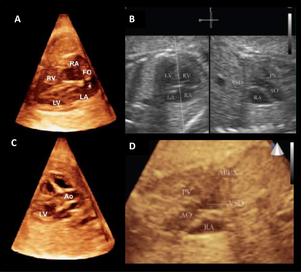
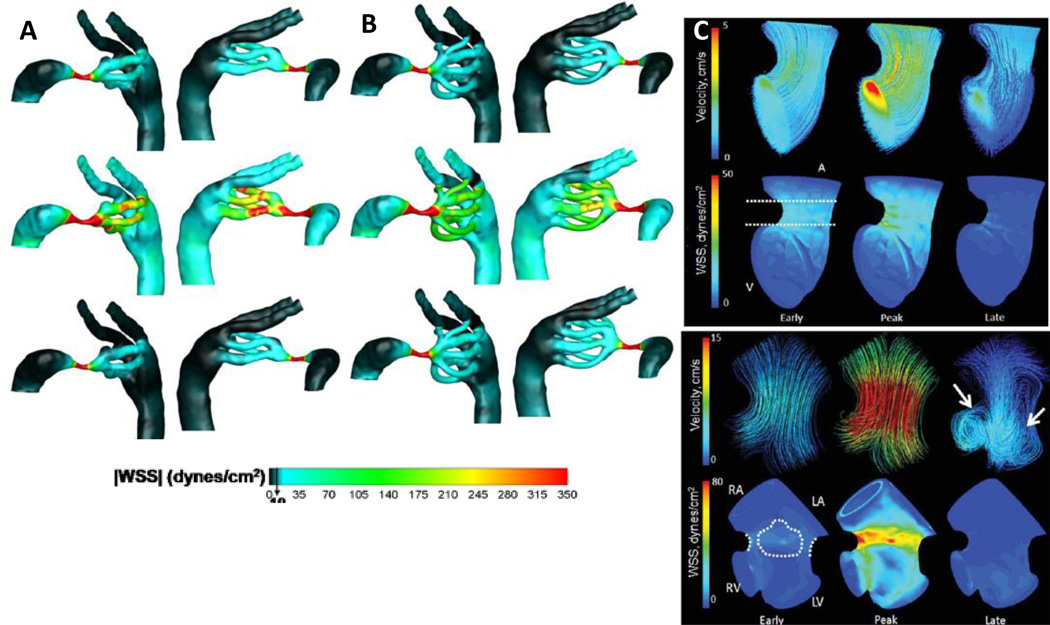
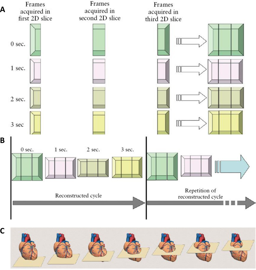
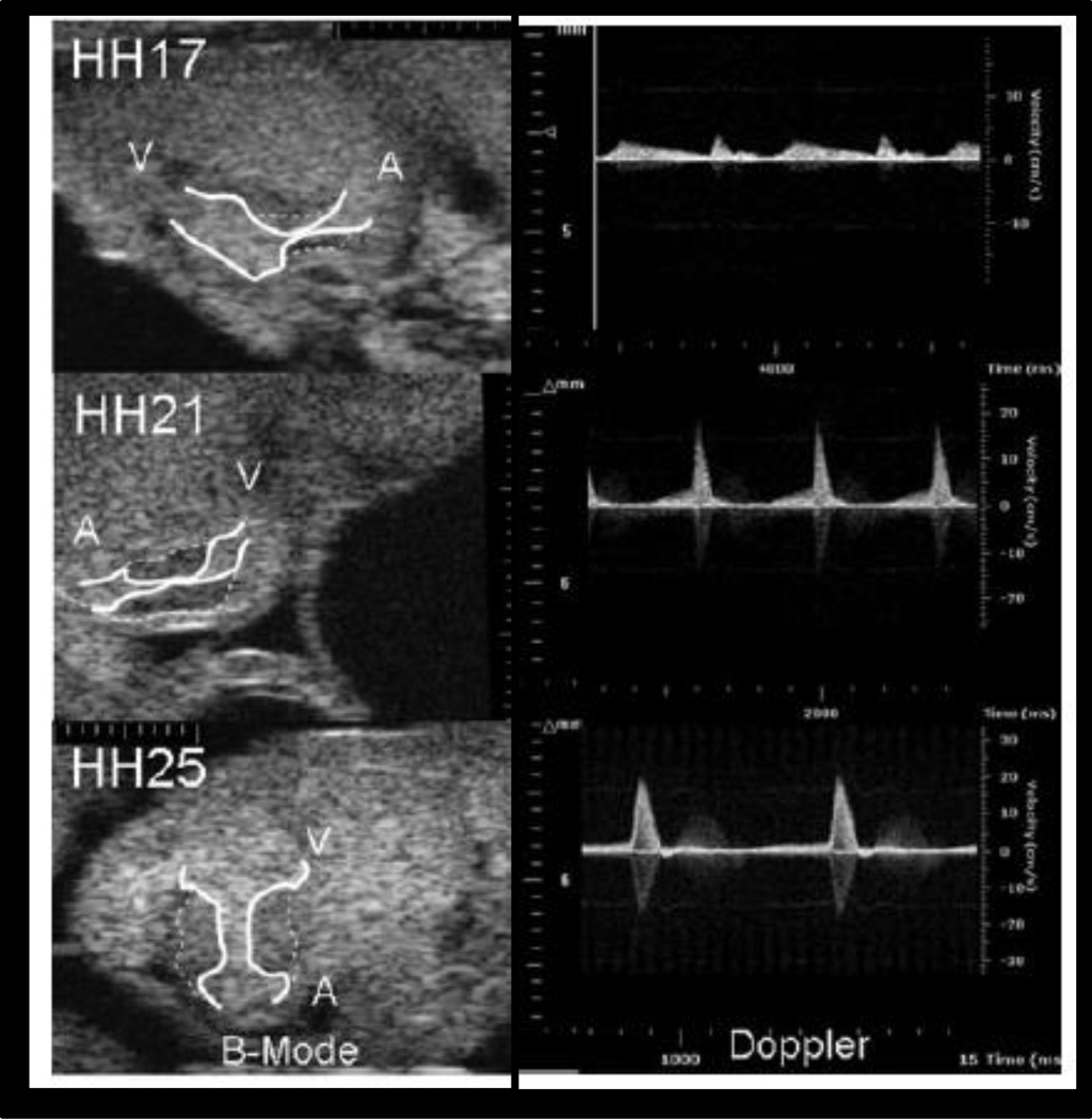
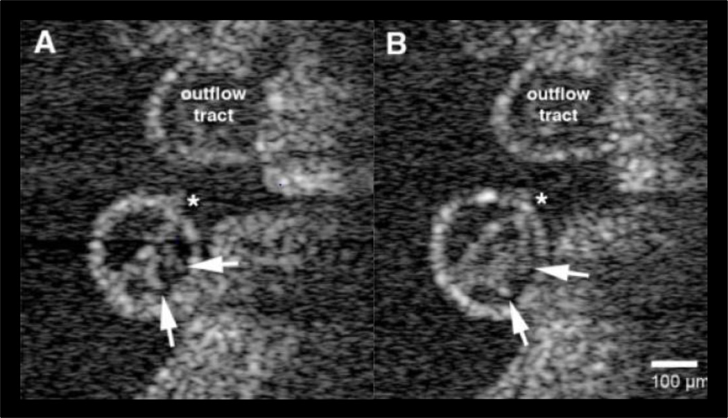
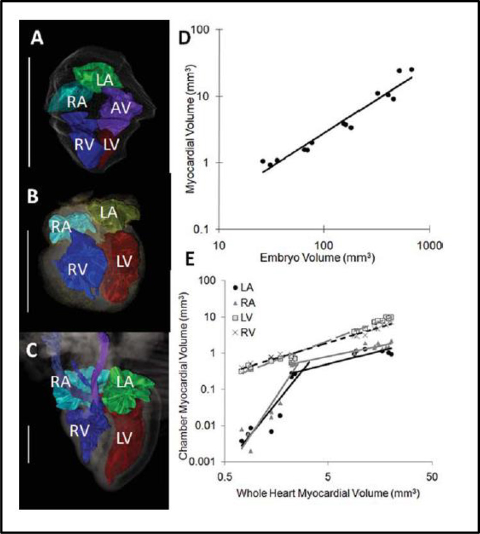
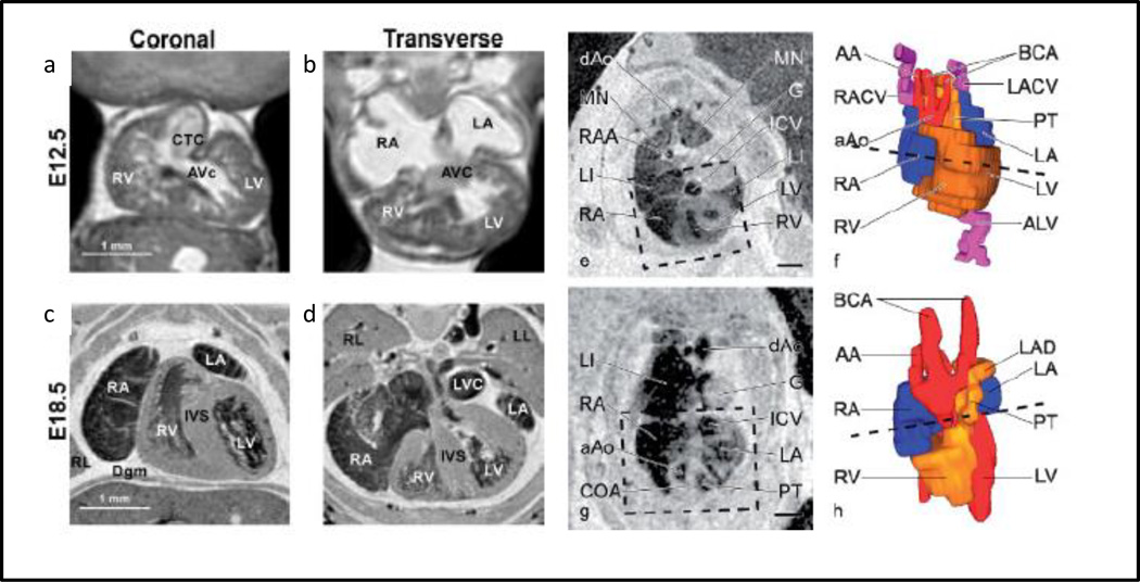
Congenital heart defects (CHD) are the most prevalent congenital disease, with 45% of deaths resulting from a congenital defect due to a cardiac malformation. Clinically significant CHD permit survival upon birth, but may become immediately life threatening. Advances in surgical intervention have significantly reduced perinatal mortality, but the outcome for many malformations is bleak. Furthermore, patients living while tolerating a CHD often acquire additional complications due to the long-term systemic blood flow changes caused by even subtle anatomical abnormalities. Accurate diagnosis of defects during fetal development is critical for interventional planning and improving patient outcomes. Advances in quantitative, multidimensional imaging are necessary to uncover the basic scientific and clinically relevant morphogenetic changes and associated hemodynamic consequences influencing normal and abnormal heart development. Ultrasound is the most widely used clinical imaging technology for assessing fetal cardiac development. Ultrasound-based fetal assessment modalities include motion mode (M-mode), two dimensional (2D), and 3D/4D imaging. These datasets can be combined with computational fluid dynamics analysis to yield quantitative, volumetric, and physiological data. Additional imaging modalities, however, are available to study basic mechanisms of cardiogenesis, including optical coherence tomography, microcomputed tomography, and magnetic resonance imaging. Each imaging technology has its advantages and disadvantages regarding resolution, depth of penetration, soft tissue contrast considerations, and cost. In this review, we analyze the current clinical and scientific imaging technologies, research studies utilizing them, and appropriate animal models reflecting clinically relevant cardiogenesis and cardiac malformations. We conclude with discussing the translational impact and future opportunities for cardiovascular development imaging research.

摘要

先天性心脏病(CHD)是最常见的先天性疾病,45%的先天性缺陷导致的死亡是由心脏畸形引起的。具有临床意义的CHD患儿出生后可存活,但可能立即危及生命。外科手术干预的进展显著降低了围产期死亡率,但许多畸形的预后仍然不容乐观。此外,患有CHD并存活下来的患者,由于即使是细微的解剖异常所导致的长期全身血流变化,往往会出现其他并发症。在胎儿发育期间准确诊断缺陷对于干预计划和改善患者预后至关重要。定量、多维成像技术的进步对于揭示影响正常和异常心脏发育的基本科学及临床相关形态发生变化以及相关血流动力学后果是必要的。超声是评估胎儿心脏发育最广泛使用的临床成像技术。基于超声的胎儿评估方式包括运动模式(M型)、二维(2D)和三维/四维成像。这些数据集可以与计算流体动力学分析相结合,以产生定量、容积和生理数据。然而,还有其他成像方式可用于研究心脏发生的基本机制,包括光学相干断层扫描、微型计算机断层扫描和磁共振成像。每种成像技术在分辨率、穿透深度、软组织对比度考量和成本方面都有其优缺点。在本综述中,我们分析了当前的临床和科学成像技术、利用这些技术的研究以及反映临床相关心脏发生和心脏畸形的合适动物模型。我们最后讨论了心血管发育成像研究的转化影响和未来机遇。

https://cdn.ncbi.nlm.nih.gov/pmc/blobs/dfd8/4472488/ffaac2c3b402/nihms694404f7.jpg
https://cdn.ncbi.nlm.nih.gov/pmc/blobs/dfd8/4472488/dd56db831bd7/nihms694404f1.jpg
https://cdn.ncbi.nlm.nih.gov/pmc/blobs/dfd8/4472488/9953abdaa078/nihms694404f2.jpg
https://cdn.ncbi.nlm.nih.gov/pmc/blobs/dfd8/4472488/341a417a7f48/nihms694404f3.jpg
https://cdn.ncbi.nlm.nih.gov/pmc/blobs/dfd8/4472488/a44f384c8fbf/nihms694404f4.jpg
https://cdn.ncbi.nlm.nih.gov/pmc/blobs/dfd8/4472488/78ed2c2accb9/nihms694404f5.jpg
https://cdn.ncbi.nlm.nih.gov/pmc/blobs/dfd8/4472488/3d4d390f7930/nihms694404f6.jpg
https://cdn.ncbi.nlm.nih.gov/pmc/blobs/dfd8/4472488/ffaac2c3b402/nihms694404f7.jpg

相似文献

1
Translational paradigms in scientific and clinical imaging of cardiac development.心脏发育的科学与临床成像中的转化范式。
Birth Defects Res C Embryo Today. 2013 Jun;99(2):106-20. doi: 10.1002/bdrc.21034.
2
Phenotyping cardiac and structural birth defects in fetal and newborn mice.在胎儿和新生鼠中表型分析心脏和结构出生缺陷。
Birth Defects Res. 2017 Jun 1;109(10):778-790. doi: 10.1002/bdr2.1048. Epub 2017 May 22.
3
Advances in fetal cardiac imaging.胎儿心脏成像的进展。
Pediatr Cardiol. 2004 May-Jun;25(3):307-21. doi: 10.1007/s00246-003-0594-0.
4
Imaging techniques for visualizing and phenotyping congenital heart defects in murine models.用于可视化和表型分析小鼠模型先天性心脏缺陷的成像技术。
Birth Defects Res C Embryo Today. 2013 Jun;99(2):93-105. doi: 10.1002/bdrc.21037.
5
A review of the complementary information available with cardiac magnetic resonance imaging and multi-slice computed tomography (CT) during the study of congenital heart disease.先天性心脏病研究中,心脏磁共振成像与多层计算机断层扫描(CT)的补充信息综述。
Int J Cardiovasc Imaging. 2004 Dec;20(6):569-78. doi: 10.1007/s10554-004-7021-3.
6
Assessment of Fetal Congenital Heart Diseases by 4-Dimensional Ultrasound Using Spatiotemporal Image Correlation: Pictorial Review.使用时空图像相关技术的四维超声评估胎儿先天性心脏病:图谱综述
Ultrasound Q. 2018 Mar;34(1):11-17. doi: 10.1097/RUQ.0000000000000328.
7
Prevalence of prenatal brain abnormalities in fetuses with congenital heart disease: a systematic review.先天性心脏病胎儿产前脑异常的患病率:一项系统评价。
Ultrasound Obstet Gynecol. 2016 Sep;48(3):296-307. doi: 10.1002/uog.15932.
8
Three-dimensional and four-dimensional flow assessment in congenital heart disease.先天性心脏病的三维和四维血流评估。
Heart. 2020 Mar;106(6):421-426. doi: 10.1136/heartjnl-2019-315797. Epub 2019 Dec 19.
9
Diagnostic quality of 3Tesla postmortem magnetic resonance imaging in fetuses with and without congenital heart disease.3T 磁共振成像在有和无先天性心脏病胎儿中的诊断质量。
Am J Obstet Gynecol. 2021 Aug;225(2):189.e1-189.e30. doi: 10.1016/j.ajog.2021.02.030. Epub 2021 Mar 1.
10
Added value of three-/four-dimensional ultrasound in offline analysis and diagnosis of congenital heart disease.三维/四维超声在先天性心脏病离线分析诊断中的附加价值。
Ultrasound Obstet Gynecol. 2011 Apr;37(4):432-7. doi: 10.1002/uog.8867. Epub 2011 Mar 7.

引用本文的文献

1
Comparative analysis of metallic nanoparticles as exogenous soft tissue contrast for live in vivo micro-computed tomography imaging of avian embryonic morphogenesis.金属纳米颗粒作为外源性软组织对比剂用于鸡胚形态发生的活体体内微型计算机断层扫描成像的比较分析。
Dev Dyn. 2016 Oct;245(10):1001-10. doi: 10.1002/dvdy.24433. Epub 2016 Aug 18.
2
Respiratory Effects on Fontan Circulation During Rest and Exercise Using Real-Time Cardiac Magnetic Resonance Imaging.使用实时心脏磁共振成像对静息和运动期间Fontan循环的呼吸影响
Ann Thorac Surg. 2016 May;101(5):1818-25. doi: 10.1016/j.athoracsur.2015.11.011. Epub 2016 Feb 10.

本文引用的文献

1
A series of normal stages in the development of the chick embryo.鸡胚胎发育的一系列正常阶段。
J Morphol. 1951 Jan;88(1):49-92.
2
Critical transitions in early embryonic aortic arch patterning and hemodynamics.早期胚胎主动脉弓模式形成和血液动力学的关键转变。
PLoS One. 2013;8(3):e60271. doi: 10.1371/journal.pone.0060271. Epub 2013 Mar 21.
3
Transgenic quail as a model for research in the avian nervous system: a comparative study of the auditory brainstem.转基因鹌鹑作为禽类神经系统研究模型:听觉脑干的比较研究。
J Comp Neurol. 2013 Jan 1;521(1):5-23. doi: 10.1002/cne.23187.
4
Fetal cardiac function: M-mode and 4D spatiotemporal image correlation.胎儿心脏功能:M 模式和 4D 时空图像相关。
Fetal Diagn Ther. 2012;32(1-2):17-21. doi: 10.1159/000335357. Epub 2012 Jul 6.
5
Quantitative in vivo imaging of embryonic development: opportunities and challenges.胚胎发育的定量活体成像:机遇与挑战。
Differentiation. 2012 Jul;84(1):149-62. doi: 10.1016/j.diff.2012.05.003. Epub 2012 Jun 12.
6
Comparison of real-time three-dimensional echocardiography and spatiotemporal image correlation in assessment of fetal interventricular septum.实时三维超声心动图与时空图像相关技术在评估胎儿室间隔中的比较。
J Matern Fetal Neonatal Med. 2012 Nov;25(11):2333-8. doi: 10.3109/14767058.2012.695822. Epub 2012 Jun 13.
7
Novel application of four-dimensional sonography with B-flow imaging and spatiotemporal image correlation in the assessment of fetal congenital heart defects.四维超声联合B-flow成像及时空关联成像在胎儿先天性心脏缺陷评估中的新应用
Echocardiography. 2012 May;29(5):614-9. doi: 10.1111/j.1540-8175.2011.01639.x. Epub 2012 Mar 8.
8
Computational hemodynamic optimization predicts dominant aortic arch selection is driven by embryonic outflow tract orientation in the chick embryo.计算血流动力学优化预测主导主动脉弓的选择是由鸡胚胚胎流出道的方向决定的。
Biomech Model Mechanobiol. 2012 Sep;11(7):1057-73. doi: 10.1007/s10237-012-0373-z. Epub 2012 Feb 4.
9
Increasing the field-of-view of dynamic cardiac OCT via post-acquisition mosaicing without affecting frame-rate or spatial resolution.通过采集后拼接增加动态心脏光学相干断层扫描的视野,同时不影响帧率或空间分辨率。
Biomed Opt Express. 2011 Sep 1;2(9):2614-22. doi: 10.1364/BOE.2.002614. Epub 2011 Aug 16.
10
Imaging of patients with congenital heart disease.先天性心脏病患者的影像学检查。
Nat Rev Cardiol. 2011 Nov 1;9(2):101-15. doi: 10.1038/nrcardio.2011.162.