Department of Bio-Medical Sciences, Human Anatomy and Histology Section, University of Catania, Catania 95123, Italy.
Int J Mol Sci. 2013 Jul 29;14(8):15767-84. doi: 10.3390/ijms140815767.
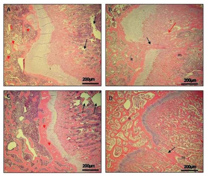
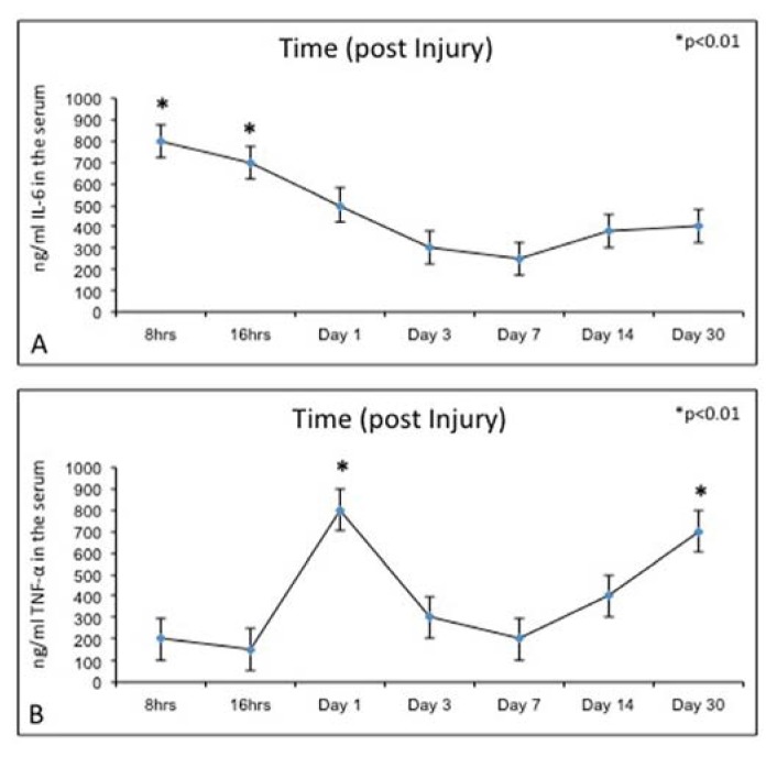
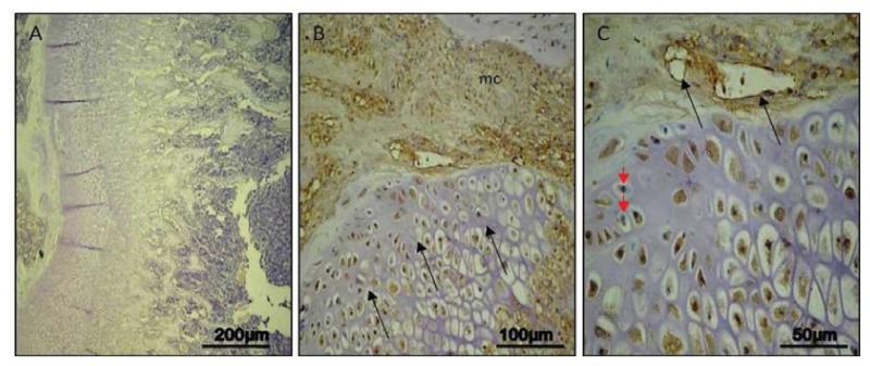
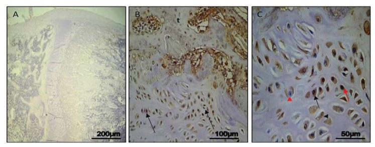
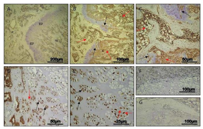
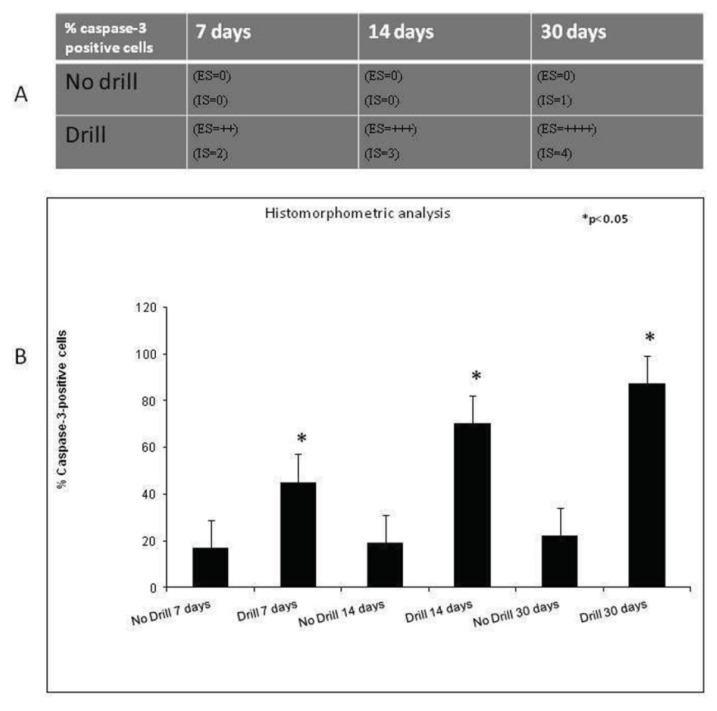
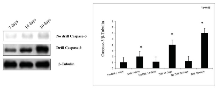
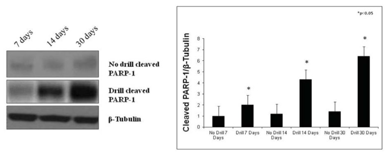
The epiphyseal plate is a hyaline cartilage plate that sits between the diaphysis and the epiphysis. The objective of this study was to determine the impact of an injury in the growth plate chondrocytes through the study of histological morphology, immunohistochemistry, histomorphometry and Western Blot analyses of the caspase-3 and cleaved PARP-1, and levels of the inflammatory cytokines, Interleukin-6 (IL-6) and Tumor Necrosis Factor alpha (TNF-α), in order to acquire more information about post-injury reactions of physeal cell turnover. In our results, morphological analysis showed that in experimental bones, neo-formed bone trabeculae-resulting from bone formation repair-invaded the growth plate and reached the metaphyseal bone tissue (bone bridge), and this could result in some growth arrest. We demonstrated, by ELISA, increased expression levels of the inflammatory cytokines IL-6 and TNF-α. Immunohistochemistry, histomorphometry and Western Blot analyses of the caspase-3 and cleaved PARP-1 showed that the physeal apoptosis rate of the experimental bones was significantly higher than that of the control ones. In conclusion, we could assume that the inflammation process causes stress to chondrocytes that will die as a biological defense mechanism, and will also increase the survival of new chondrocytes for maintaining cell homeostasis. Nevertheless, the exact stimulus leading to the increased apoptosis rate, observed after injury, needs additional research to understand the possible contribution of chondrocyte apoptosis to growth disturbance.
骺板是位于骨干和骺之间的透明软骨板。本研究的目的是通过研究生长板软骨细胞的组织形态学、免疫组织化学、组织形态计量学和 caspase-3 和裂解 PARP-1 的 Western Blot 分析,以及白细胞介素 6 (IL-6) 和肿瘤坏死因子 α (TNF-α) 等炎症细胞因子的水平,来确定生长板软骨细胞损伤对骺板的影响,从而获得更多关于骺细胞损伤后反应的信息。在我们的研究结果中,形态学分析表明,在实验骨中,新形成的骨小梁(源于骨形成修复)侵入生长板并到达骺端骨组织(骨桥),这可能导致生长停滞。通过 ELISA 我们证明了炎症细胞因子 IL-6 和 TNF-α的表达水平增加。免疫组织化学、组织形态计量学和 caspase-3 和裂解 PARP-1 的 Western Blot 分析表明,实验骨的骺板细胞凋亡率明显高于对照组。总之,我们可以假设炎症过程会对软骨细胞造成压力,作为一种生物防御机制,软骨细胞会死亡,并增加新的软骨细胞的存活以维持细胞内稳态。然而,对于导致损伤后细胞凋亡率增加的确切刺激,还需要进一步的研究来了解软骨细胞凋亡对生长障碍的可能贡献。