Department of Radiology, Chonnam National University Medical School, Chonnam National University Hwasun Hospital, Hwasun 519-763, Korea.
Korean J Radiol. 2013 Jul-Aug;14(4):551-8. doi: 10.3348/kjr.2013.14.4.551. Epub 2013 Jul 17.
To analyze the magnetic resonance (MR) imaging findings of invasive micropapillary carcinoma of the breast.
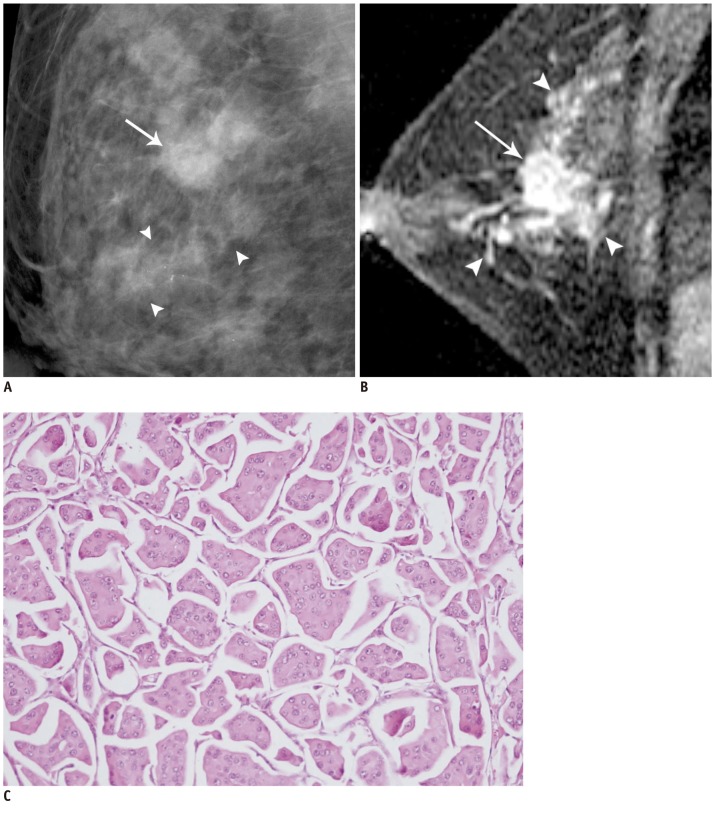
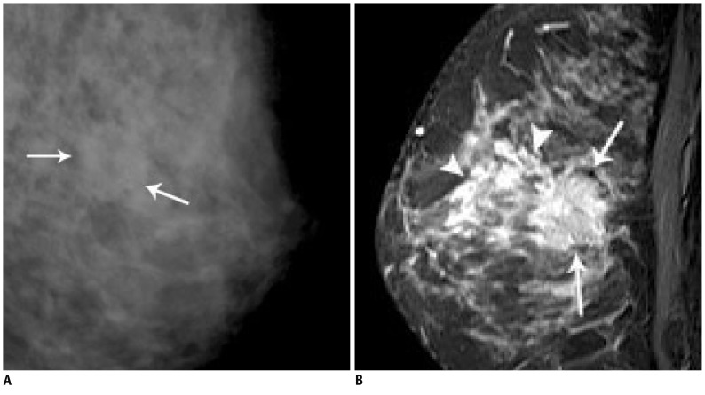
MR images were retrospectively evaluated in 14 patients (age range: 37-67, mean age: 49 years) with pathologically confirmed invasive micropapillary carcinoma of the breast. The enhancement type (mass/non-mass), shape, margin, contrast enhancement, and time-intensity curve pattern on the dynamic study were correlated with the histopathologic features. Associated findings, such as edema, nipple change, skin change and enlarged axillary lymph nodes were also studied.
The most common features of the masses were irregular shape (12 of 14 patients, 85.8%) and irregular or spiculated margin (11 of 14 patients, 78.7%). The contrast enhancement was heterogeneous in 11 patients (78.7%), rim enhancement in 2 cases (14.2%), and homogeneous in one patient (7.1%). The predominant kinetic pattern was rapid increase (14 of 14, 100%) in the initial phase and washout (11 of 14, 78.7%) in the delayed phase. Associated non-mass like enhancement was shown in 4 patients, representing ductal carcinoma in situ. MR imaging helped detect additional sites of cancer other than the index lesion in 3 patients (21.4%). Enlarged axillary lymphadenopathy was identified in 7 of the 14 patients (50%).
Invasive micropapillary carcinoma appears as a mass with an irregular shape, irregular or spiculated margin and heterogeneous enhancement on MR imaging. Though these findings are not specific and are also observed with other breast malignancies, invasive micropapillary carcinoma frequently showed multiple lesions, accompanying non-mass enhancement and axillary lymph node enlargement.
分析乳腺浸润性微乳头状癌的磁共振(MR)成像表现。
回顾性分析 14 例经病理证实为乳腺浸润性微乳头状癌患者的 MR 图像(年龄范围:37-67 岁,平均年龄:49 岁)。将增强类型(肿块/非肿块)、形态、边缘、对比增强及动态研究的时间-强度曲线模式与组织病理学特征相关联。还研究了相关发现,如水肿、乳头改变、皮肤改变和腋窝淋巴结肿大。
肿块的最常见特征是不规则形状(14 例患者中的 12 例,85.8%)和不规则或锯齿状边缘(14 例患者中的 11 例,78.7%)。11 例患者(78.7%)的对比增强呈不均匀,2 例(14.2%)呈边缘增强,1 例(7.1%)呈均匀增强。主要的动力学模式是在初始阶段快速增加(14 例患者中的 14 例,100%)和在延迟阶段洗脱(14 例患者中的 11 例,78.7%)。4 例患者显示非肿块样增强,代表导管原位癌。MR 成像有助于在 3 例患者(21.4%)中发现除了指数病变之外的其他癌症部位。14 例患者中有 7 例(50%)发现腋窝淋巴结肿大。
乳腺浸润性微乳头状癌在 MR 成像上表现为形态不规则、边缘不规则或锯齿状、增强不均匀的肿块。虽然这些发现并不特异,也见于其他乳腺恶性肿瘤,但浸润性微乳头状癌常表现为多发病灶,伴有非肿块样增强和腋窝淋巴结肿大。