Lee Gwang-Jun, Jung Tae-Young, Choi Seong-Min, Jung Min-Young
Department of Neurosurgery, Chonnam National University Research Institute of Medical Sciences, Chonnam National University Hwasun Hospital & Medical School, Gwangju, Korea.
J Korean Neurosurg Soc. 2013 May;53(5):312-5. doi: 10.3340/jkns.2013.53.5.312. Epub 2013 May 31.
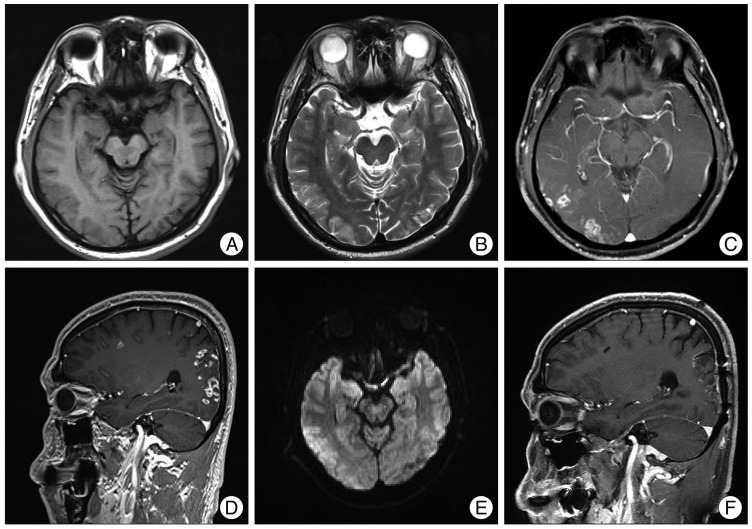
Aspergillosis in the central nervous system (CNS) is a very rare disease in immune-competent patients. There was a case of a healthy man without a history of immune-compromised disease who had invasive aspergillosis with unusual radiologic findings. A 48-year-old healthy man with diabetes mellitus, presented with complaints of blurred vision that persisted for one month. Brain magnetic resonance imaging (MRI) showed multiple nodular enhancing lesions on the right cerebral hemisphere. The diffusion image appeared in a high-signal intensity in these areas. Cerebrospinal fluid examination did not show any infection signs. An open biopsy was done and intraoperative findings showed grayish inflammatory and necrotic tissue without a definitive mass lesion. The pathologic result was a brain abscess caused by fungal infection, morphologically aspergillus. Antifungal agents (Amphotericin B, Ambisome and Voriconazole) were used for treatment for 3 months. The visual symptoms improved. There was no recurrence or abscess pocket, but the remaining focal enhanced lesions were visible in the right temporal and occipital area at a one year follow-up MRI. This immune-competent patient showed multiple enhancing CNS aspergillosis in the cerebral hemisphere, which had a good outcome with antifungal agents.
中枢神经系统曲霉病在免疫功能正常的患者中是一种非常罕见的疾病。有一例无免疫功能低下疾病史的健康男性发生了具有不寻常影像学表现的侵袭性曲霉病。一名48岁患有糖尿病的健康男性,主诉视力模糊持续了一个月。脑部磁共振成像(MRI)显示右侧大脑半球有多个结节状强化病灶。这些区域的扩散图像呈高信号强度。脑脊液检查未显示任何感染迹象。进行了开放性活检,术中发现灰白色炎性和坏死组织,无明确的肿块病变。病理结果是真菌感染引起的脑脓肿,形态学上为曲霉菌。使用抗真菌药物(两性霉素B、安必素和伏立康唑)治疗3个月。视觉症状有所改善。没有复发或脓肿腔,但在一年后的随访MRI中,右侧颞叶和枕叶区域仍可见残留的局灶性强化病灶。这名免疫功能正常的患者在大脑半球出现了多发性强化性中枢神经系统曲霉病,使用抗真菌药物治疗后预后良好。