Marzolf Guillaume, Sabou Marcela, Lannes Béatrice, Cotton François, Meyronet David, Galanaud Damien, Cottier Jean-Philippe, Grand Sylvie, Desal Hubert, Kreutz Julie, Schenck Maleka, Meyer Nicolas, Schneider Francis, Dietemann Jean-Louis, Koob Meriam, Herbrecht Raoul, Kremer Stéphane
Département de Neuroradiologie, Hôpitaux Universitaires de Strasbourg, Strasbourg, France.
Laboratoire de Parasitologie et de Mycologie Médicale, Hôpitaux Universitaires de Strasbourg, Strasbourg, France.
PLoS One. 2016 Apr 20;11(4):e0152475. doi: 10.1371/journal.pone.0152475. eCollection 2016.
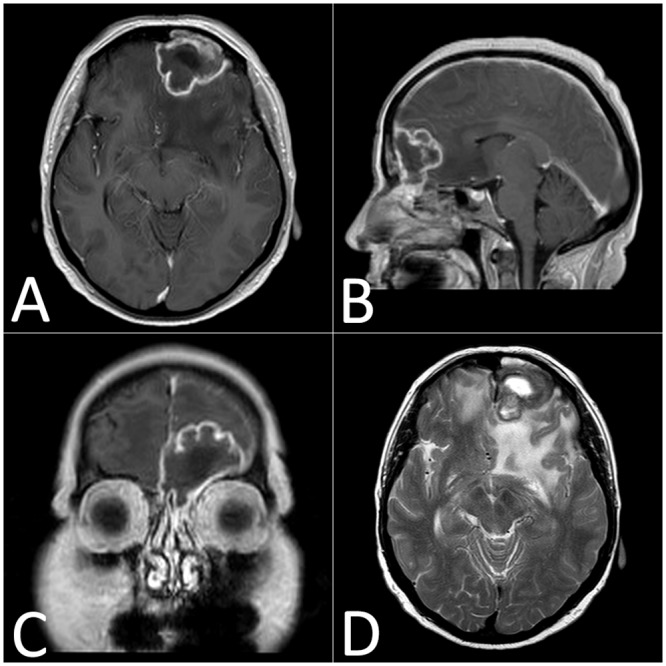
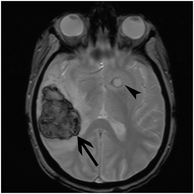
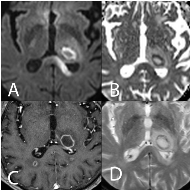
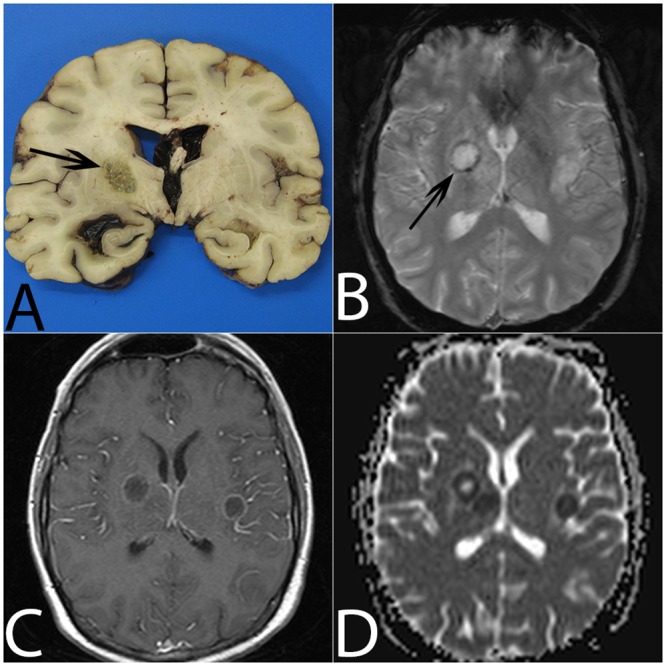
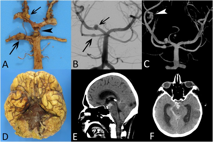
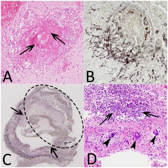
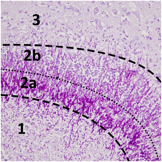
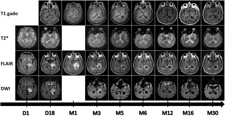
Cerebral aspergillosis is associated with a significant morbidity and mortality rate. The imaging data present different patterns and no full consensus exists on typical imaging characteristics of the cerebral lesions. We reviewed MRI findings in 21 patients with cerebral aspergillosis and correlated them to the immune status of the patients and to neuropathological findings when tissue was available. The lesions were characterized by their number, topography, and MRI signal. Dissemination to the brain resulted from direct spread from paranasal sinuses in 8 patients, 6 of them being immunocompetent. Hematogenous dissemination was observed in 13 patients, all were immunosuppressed. In this later group we identified a total of 329 parenchymal abscesses involving the whole brain with a predilection for the corticomedullary junction. More than half the patients had a corpus callosum lesion. Hemorrhagic lesions accounted for 13% and contrast enhancement was observed in 61% of the lesions. Patients with hematogenous dissemination were younger (p = 0.003), had more intracranial lesions (p = 0.0004) and had a higher 12-week mortality rate (p = 0.046) than patients with direct spread from paranasal sinuses. Analysis of 12 aneurysms allowed us to highlight two distinct situations. In case of direct spread from the paranasal sinuses, aneurysms are saccular and located on the proximal artery portions, while the hematogenous dissemination in immunocompromised patients is more frequently associated with distal and fusiform aneurysms. MRI is the exam of choice for cerebral aspergillosis. Number and type of lesions are different according to the mode of dissemination of the infection.
脑曲霉病与较高的发病率和死亡率相关。影像学数据呈现出不同的模式,对于脑病变的典型影像学特征尚无完全一致的看法。我们回顾了21例脑曲霉病患者的MRI表现,并将其与患者的免疫状态以及有组织样本时的神经病理学结果进行关联。病变通过数量、部位和MRI信号进行特征描述。8例患者的脑部感染是由鼻窦直接蔓延所致,其中6例免疫功能正常。13例患者观察到血行播散,均为免疫抑制患者。在这后一组中,我们共识别出329个脑实质脓肿,累及全脑,以皮质髓质交界区最为常见。超过半数患者有胼胝体病变。出血性病变占13%,61%的病变有强化表现。血行播散的患者比鼻窦直接蔓延的患者更年轻(p = 0.003),颅内病变更多(p = 0.0004),12周死亡率更高(p = 0.046)。对12个动脉瘤的分析使我们能够突出两种不同情况。鼻窦直接蔓延时,动脉瘤呈囊状,位于动脉近端部分,而免疫功能低下患者的血行播散更常与远端梭形动脉瘤相关。MRI是脑曲霉病的首选检查方法。根据感染的播散方式,病变的数量和类型有所不同。