Institute of Plant Physiology, Justus-Liebig-University Giessen, Germany ; Institute of General Botany, Plant Cell Biology Research Group, Justus-Liebig-University Giessen, Germany.
Front Plant Sci. 2013 Jul 31;4:274. doi: 10.3389/fpls.2013.00274. eCollection 2013.
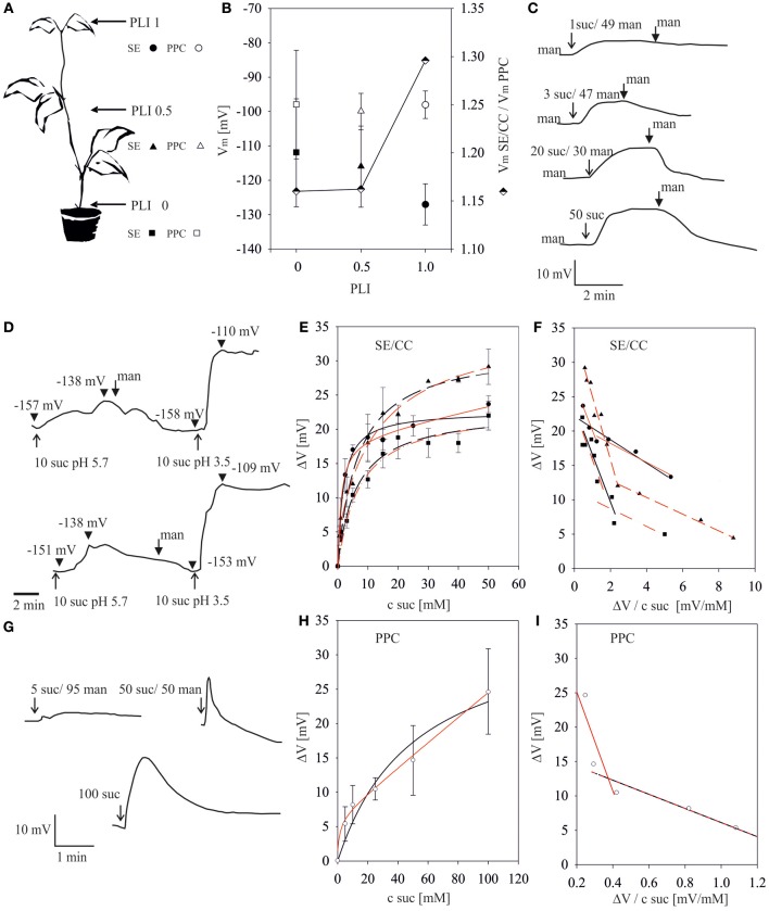
Apart from cut aphid stylets in combination with electrophysiology, no attempts have been made thus far to measure in vivo sucrose-uptake properties of sieve elements. We investigated the kinetics of sucrose uptake by single sieve elements and phloem parenchyma cells in Vicia faba plants. To this end, microelectrodes were inserted into free-lying phloem cells in the main vein of the youngest fully-expanded leaf, half-way along the stem, in the transition zone between the autotrophic and heterotrophic part of the stem, and in the root axis. A top-to-bottom membrane potential gradient of sieve elements was observed along the stem (-130 mV to -110 mV), while the membrane potential of the phloem parenchyma cells was stable (approx. -100 mV). In roots, the membrane potential of sieve elements dropped abruptly to -55 mV. Bathing solutions having various sucrose concentrations were administered and sucrose/H(+)-induced depolarizations were recorded. Data analysis by non-linear least-square data fittings as well as by linear Eadie-Hofstee (EH) -transformations pointed at biphasic Michaelis-Menten kinetics (2 MM, EH: K m1 1.2-1.8 mM, K m2 6.6-9.0 mM) of sucrose uptake by sieve elements. However, Akaike's Information Criterion (AIC) favored single MM kinetics. Using single MM as the best-fitting model, K m values for sucrose uptake by sieve elements decreased along the plant axis from 1 to 7 mM. For phloem parenchyma cells, higher K m values (EH: K m1 10 mM, K m2 70 mM) as compared to sieve elements were found. In preliminary patch-clamp experiments with sieve-element protoplasts, small sucrose-coupled proton currents (-0.1 to -0.3 pA/pF) were detected in the whole-cell mode. In conclusion (a) K m values for sucrose uptake measured by electrophysiology are similar to those obtained with heterologous systems, (b) electrophysiology provides a useful tool for in situ determination of K m values, (c) As yet, it remains unclear if one or two uptake systems are involved in sucrose uptake by sieve elements, (d) Affinity for sucrose uptake by sieve elements exceeds by far that by phloem parenchyma cells, (e) Patch-clamp studies provide a feasible basis for quantification of sucrose uptake by single cells. The consequences of the findings for whole-plant carbohydrate partitioning are discussed.
除了切下蚜虫口针并结合电生理学外,迄今为止,尚未尝试测量筛管中单筛管和韧皮部薄壁细胞的活体蔗糖吸收特性。我们研究了 Vicia faba 植物中单筛管和韧皮部薄壁细胞的蔗糖吸收动力学。为此,将微电极插入主叶脉中自由悬浮的韧皮部细胞、茎中部、茎自养和异养部分之间的过渡区以及根轴中。沿茎观察到筛管的顶端到底部的膜电位梯度(-130 mV 至-110 mV),而韧皮部薄壁细胞的膜电位稳定(约-100 mV)。在根中,筛管的膜电位突然降至-55 mV。施用具有不同蔗糖浓度的浴液,并记录蔗糖/H(+)诱导的去极化。通过非线性最小二乘数据拟合以及线性 Eadie-Hofstee(EH)-变换进行数据分析表明,蔗糖吸收具有双相米氏-门登动力学(2 MM,EH:K m1 为 1.2-1.8 mM,K m2 为 6.6-9.0 mM)。然而,Akaike 的信息准则(AIC)支持单 MM 动力学。使用单 MM 作为最佳拟合模型,沿植物轴,筛管对蔗糖的 K m 值从 1 到 7 mM 降低。对于韧皮部薄壁细胞,与筛管相比,发现了更高的 K m 值(EH:K m1 为 10 mM,K m2 为 70 mM)。在筛管原生质体的初步膜片钳实验中,在全细胞模式下检测到小的蔗糖偶联质子电流(-0.1 至-0.3 pA/pF)。总之,(a)电生理学测量的蔗糖吸收 K m 值与异源系统获得的值相似,(b)电生理学为原位测定 K m 值提供了有用的工具,(c)目前尚不清楚是否涉及一个或两个摄取系统参与筛管中蔗糖的摄取,(d)筛管对蔗糖的亲和力远远超过韧皮部薄壁细胞,(e)膜片钳研究为量化单细胞蔗糖摄取提供了可行的基础。讨论了这些发现对整个植物碳水化合物分配的影响。