Lee Jang-Hoon, Do Hyung-Dong, Lee Jung-Cheul
J Cardiothorac Surg. 2013 Aug 6;8:181. doi: 10.1186/1749-8090-8-181.
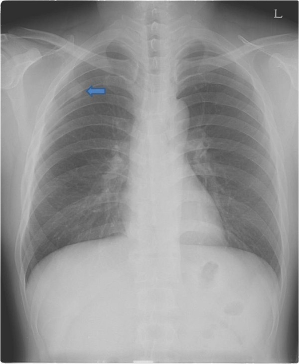
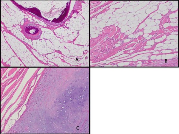
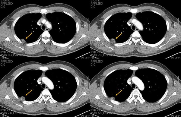
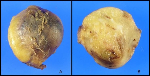
A tumor shadow was identified in the chest X-ray of a 40-year-old Korean man and he was referred to our hospital. The computed tomographic (CT) scan of his chest showed a 3-cm rounded pleural-based mass lesion with calcification, which was growing into the intercostal muscles. Thoracoscopic surgery was performed to resect the tumor. From the histological findings, the tumor was diagnosed as an intramuscular lipoma. The patient displayed no evidence of recurrence for more than 18 months. As well-circumscribed type of intramuscular lipoma is a rare tumor, we report this case with a literature review in this paper.
一名40岁的韩国男性胸部X光检查发现有肿瘤阴影,随后转诊至我院。其胸部计算机断层扫描(CT)显示一个3厘米大小、圆形、位于胸膜的肿块病变,伴有钙化,且已侵入肋间肌。遂行胸腔镜手术切除肿瘤。根据组织学检查结果,该肿瘤被诊断为肌内脂肪瘤。患者术后18个月以上无复发迹象。由于边界清晰型肌内脂肪瘤是一种罕见肿瘤,我们在此报告该病例并进行文献复习。