Department of Ophthalmology, Kagoshima University Graduate School of Medical and Dental Sciences, Kagoshima, Japan.
PLoS One. 2013 Jul 29;8(7):e69994. doi: 10.1371/journal.pone.0069994. Print 2013.
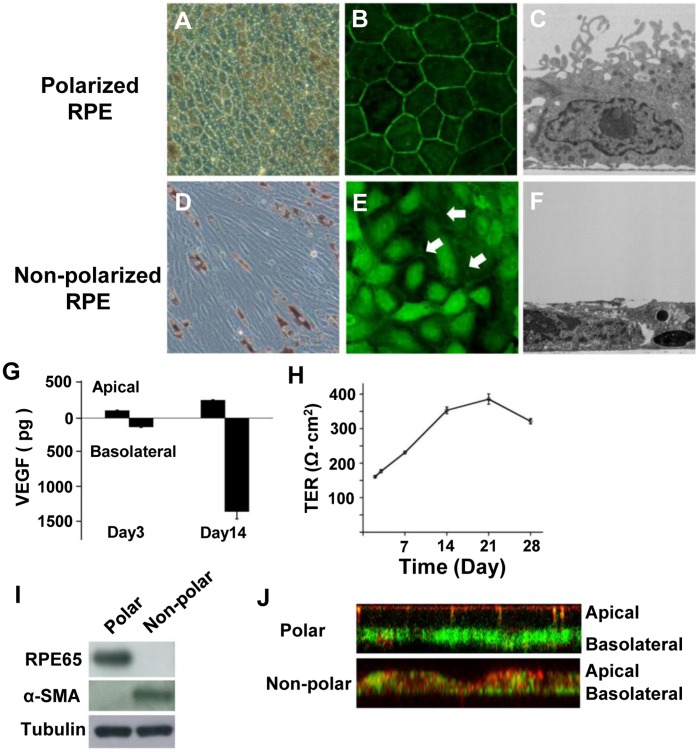
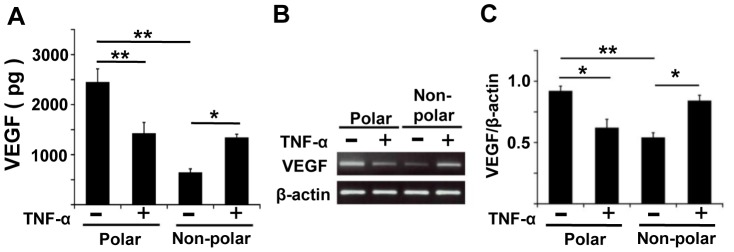
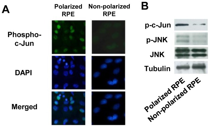
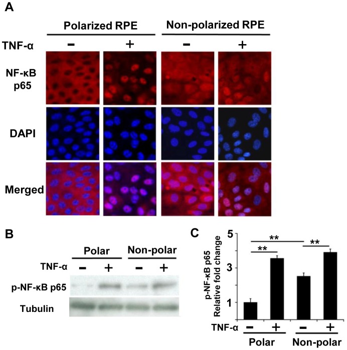
Asymmetrical secretion of vascular endothelial growth factor (VEGF) by retinal pigment epithelial (RPE) cells in situ is critical for maintaining the homeostasis of the retina and choroid. VEGF is also involved in the development and progression of age-related macular degeneration (AMD). We studied the effect of tumor necrosis factor-α (TNF-α) on the secretion of VEGF in polarized and non-polarized RPE cells (P-RPE cells and N-RPE cells, respectively) in culture and in situ in rats. A subretinal injection of TNF-α caused a decrease in VEGF expression and choroidal atrophy. Porcine RPE cells were seeded on Transwell™ filters, and their maturation and polarization were confirmed by the asymmetrical VEGF secretion and trans electrical resistance. Exposure to TNF-α decreased the VEGF secretion in P-RPE cells but increased it in N-RPE cells in culture. TNF-α inactivated JNK in P-RPE cells but activated it in N-RPE cells, and TNF-α activated NF-κB in P-RPE cells but not in N-RPE cells. Inhibition of NF-κB activated JNK in both types of RPE cells indicating crosstalk between JNK and NF-κB. TNF-α induced the inhibitory effects of NF-κB on JNK in P-RPE cells because NF-κB is continuously inactivated. In N-RPE cells, however, it was not evident because NF-κB was already activated. The basic activation pattern of JNK and NF-κB and their crosstalk led to opposing responses of RPE cells to TNF-α. These results suggest that VEGF secretion under inflammatory conditions depends on cellular polarization, and the TNF-α-induced VEGF down-regulation may result in choroidal atrophy in polarized physiological RPE cells. TNF-α-induced VEGF up-regulation may cause neovascularization by non-polarized or non-physiological RPE cells.
血管内皮生长因子(VEGF)在视网膜色素上皮(RPE)细胞中的不对称分泌对于维持视网膜和脉络膜的内稳态至关重要。VEGF 还参与年龄相关性黄斑变性(AMD)的发展和进展。我们研究了肿瘤坏死因子-α(TNF-α)对培养中和大鼠体内极化和非极化 RPE 细胞(分别为 P-RPE 细胞和 N-RPE 细胞)中 VEGF 分泌的影响。眼后注射 TNF-α可导致 VEGF 表达减少和脉络膜萎缩。将猪 RPE 细胞接种在 Transwell™过滤器上,通过不对称 VEGF 分泌和跨膜电阻证实其成熟和极化。暴露于 TNF-α可减少培养中 P-RPE 细胞中的 VEGF 分泌,但增加 N-RPE 细胞中的 VEGF 分泌。TNF-α在 P-RPE 细胞中使 JNK 失活,但在 N-RPE 细胞中激活 JNK,TNF-α在 P-RPE 细胞中激活 NF-κB,但在 N-RPE 细胞中不激活 NF-κB。NF-κB 抑制剂激活了两种类型的 RPE 细胞中的 JNK,表明 JNK 和 NF-κB 之间存在串扰。TNF-α诱导 P-RPE 细胞中 NF-κB 对 JNK 的抑制作用,因为 NF-κB 持续失活。然而,在 N-RPE 细胞中,由于 NF-κB 已经被激活,这种情况并不明显。JNK 和 NF-κB 的基本激活模式及其串扰导致 RPE 细胞对 TNF-α的反应相反。这些结果表明,炎症条件下的 VEGF 分泌取决于细胞极化,TNF-α诱导的 VEGF 下调可能导致极化生理 RPE 细胞中的脉络膜萎缩。TNF-α诱导的 VEGF 上调可能导致非极化或非生理 RPE 细胞的新生血管形成。