Department of Biologic and Materials Sciences, School of Dentistry, University of Michigan, 1011 N. University Avenue, Ann Arbor, MI 48109, USA.
Neuroscience. 2013 Nov 12;252:35-44. doi: 10.1016/j.neuroscience.2013.07.068. Epub 2013 Aug 8.
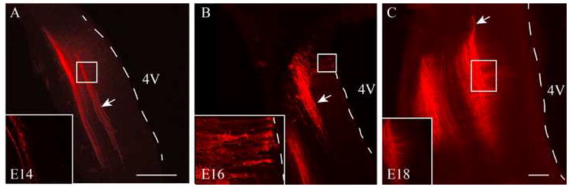
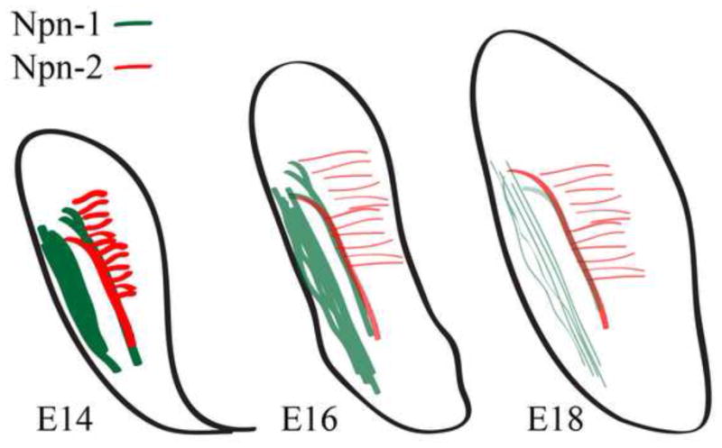
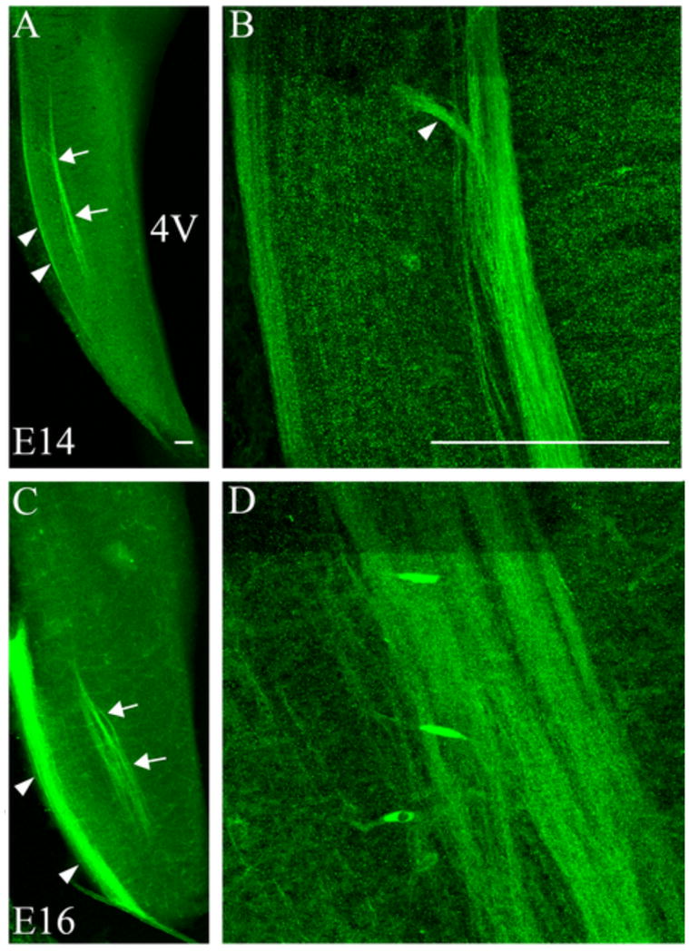
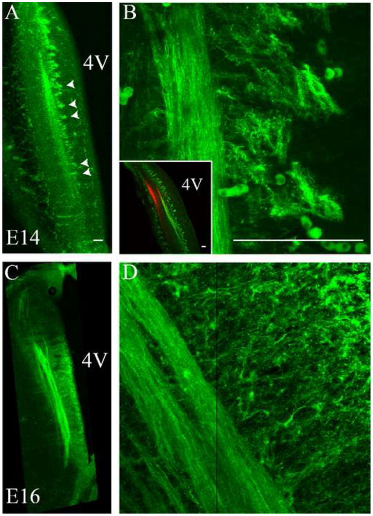
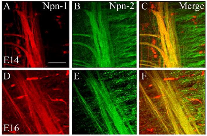
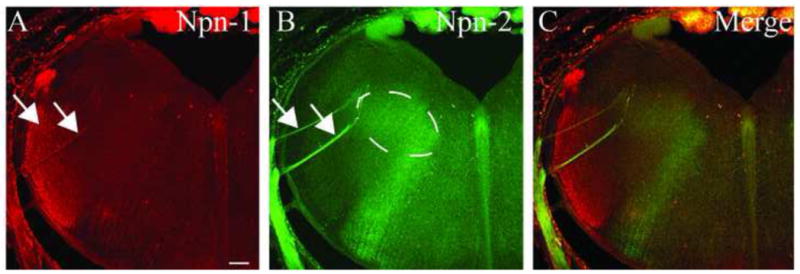
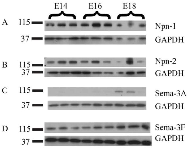
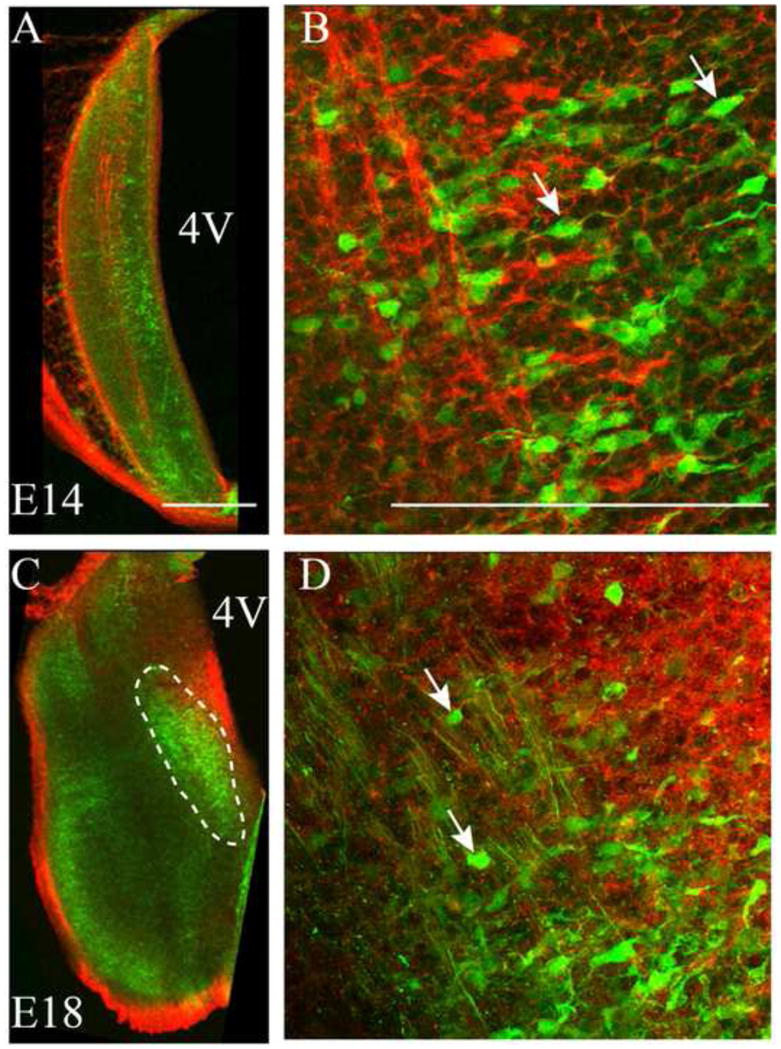
The rostral nucleus of the solitary tract (rNST) receives orosensory information from taste bud cells in the tongue and palate via cranial nerves VII and IX. These nerves enter the brainstem, form the solitary tract (ST) and synapse with neurons in the rNST, which then relay incoming sensory information to other brain areas to process external gustatory stimuli. Factors that direct or regulate the trajectory of the developing ST are largely unknown. We used 1,1'-dioctadecyl-3,3,3',3'-tetramethylindocarbocyanine perchlorate (DiI) to identify ST projections originating from cells in the geniculate ganglia of embryonic rats from embryonic day 14 through 18 (E14-E18). After identifying the ST fibers, immunolabeling for and protein expression analysis of the axon guidance molecules neuropilin-1 (Npn-1) and neuropilin-2 (Npn-2) and their binding partners, semaphorin-3A (Sema-3A) and semaphorin-3F (Sema-3F) were performed. The results detail the formation of ST projections into the gustatory brainstem and their relationship to developing rNST neurons. DiI-labeled ST fibers were present in the brainstem as early as E14. Npn-1 was expressed in the ST and in the trigeminal tract at E14, but levels of the protein declined through E18. The expression levels of the binding partner of Npn-1, Sema-3A, increased from E14 to E18. Npn-2 was expressed in the ST and, additionally, in radially oriented, tuft-like structures within the brainstem at E14. Expression levels of Npn-2 also declined through E18, in contrast to the expression levels of its binding partner, Sema-3F, which increased during this time period. For the first time, the time course and particular molecular components involved in development of the ST have been identified. These results indicate that the neuropilin and semaphorin families of axon guidance molecules are potential molecular participants in ST formation.
孤束核的吻侧部(rNST)通过颅神经 VII 和 IX 接收来自舌和 palate 味蕾的口感觉信息。这些神经进入脑干,形成孤束(ST)并与 rNST 中的神经元突触,然后将传入的感觉信息中继到其他大脑区域以处理外部味觉刺激。指导或调节发育中的 ST 轨迹的因素在很大程度上尚不清楚。我们使用 1,1'-二辛基-3,3,3',3'-四甲基吲哚羰花青高氯酸盐(DiI)来识别源自胚胎大鼠 14 至 18 天(E14-E18)的神经节神经节的 ST 投射。在识别 ST 纤维后,对轴突导向分子神经纤毛蛋白-1(Npn-1)和神经纤毛蛋白-2(Npn-2)及其结合伙伴神经调节蛋白-3A(Sema-3A)和神经调节蛋白-3F(Sema-3F)进行免疫标记和蛋白表达分析。结果详细描述了 ST 投射进入味觉脑干的形成及其与发育中的 rNST 神经元的关系。早在 E14 时,DiI 标记的 ST 纤维就存在于脑干中。Npn-1 在 E14 时在 ST 和三叉神经束中表达,但蛋白水平在 E18 时下降。Npn-1 的结合伙伴 Sema-3A 的表达水平从 E14 到 E18 增加。Npn-2 在 ST 中表达,此外,在 E14 时在脑干中呈放射状的毛状结构中表达。与它的结合伙伴 Sema-3F 的表达水平相反,Npn-2 的表达水平在这段时间内下降。这是首次确定 ST 发育中涉及的时间过程和特定分子成分。这些结果表明,神经纤毛蛋白和神经调节蛋白家族的轴突导向分子是 ST 形成的潜在分子参与者。