Dubas Ewa, Custers Jan, Kieft Henk, Wędzony Maria, van Lammeren André A M
The Franciszek Górski Institute of Plant Physiology, Polish Academy of Sciences, Niezapominajek 21, 30-239, Kraków, Poland,
Protoplasma. 2014 Jan;251(1):103-13. doi: 10.1007/s00709-013-0530-y. Epub 2013 Aug 10.
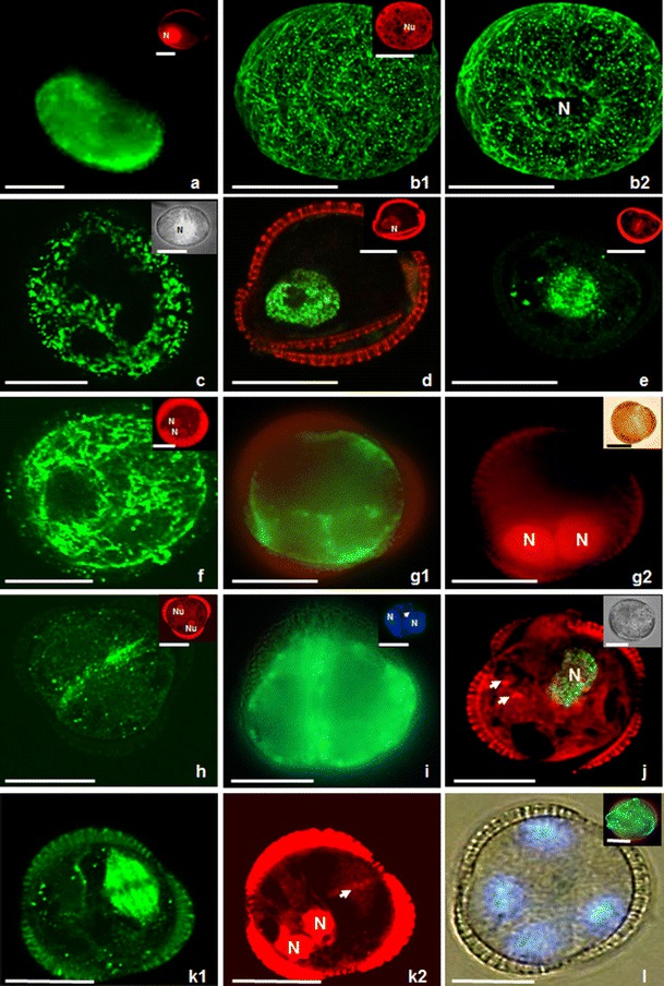
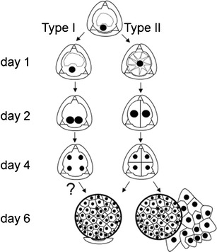
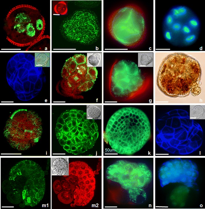
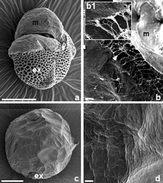
Isolated microspores of B. napus in culture change their developmental pathway from gametophytic to sporophytic and form embryo-like structures (ELS) upon prolonged heat shock treatment (5 days at 32 °C). ELS express polarity during the initial days of endosporic development. In this study, we focussed on the analysis of polarity development of ELS without suspensor. Fluorescence microscopy and 3-D confocal laser scanning microscopy (CLSM) without tissue interfering enabled us to get a good insight in the distribution of nuclei, mitochondria and endoplasmic reticulum (ER), the architecture of microtubular (MT) cytoskeleton and the places of 5-bromo-2'-deoxy-uridine (BrdU) incorporation in successive stages of microspore embryogenesis. Scanning electron microscopy (SEM) analysis revealed, for the first time, the appearance of a fibrillar extracellular matrix-like structure (ECM-like structure) in androgenic embryos without suspensor. Two types of endosporic development were distinguished based upon the initial location of the microspore nucleus. The polarity of dividing and growing cells was recognized by the differential distributions of organelles, by the organization of the MT cytoskeleton and by the visualization of DNA synthesis in the cell cycle. The directional location of nuclei, ER, mitochondria and starch grains in relation to the MTs configurations were early polarity indicators. Both exine rupture and ECM-like structure on the outer surfaces of ELS are supposed to stabilize ELS's morphological polarity. As the role of cell polarity during early endosporic microspore embryogenesis in apical-basal cell fate determination remains unclear, microspore culture system provides a powerful in vitro tool for studying the developmental processes that take place during the earliest stages of plant embryogenesis.
甘蓝型油菜离体培养的小孢子会改变其发育途径,从配子体发育途径转变为孢子体发育途径,并在长时间热激处理(32℃处理5天)后形成胚状结构(ELS)。ELS在孢子内发育的最初几天表现出极性。在本研究中,我们重点分析了无胚柄ELS的极性发育。荧光显微镜和无组织干扰的三维共聚焦激光扫描显微镜(CLSM)使我们能够深入了解细胞核、线粒体和内质网(ER)的分布、微管(MT)细胞骨架的结构以及5-溴-2'-脱氧尿苷(BrdU)在小孢子胚胎发生连续阶段的掺入位置。扫描电子显微镜(SEM)分析首次揭示了无胚柄雄核发育胚胎中出现的纤维状细胞外基质样结构(ECM样结构)。根据小孢子核的初始位置区分出两种类型的孢子内发育。通过细胞器的差异分布、MT细胞骨架的组织以及细胞周期中DNA合成的可视化来识别分裂和生长细胞的极性。细胞核、ER、线粒体和淀粉粒相对于MT构型的定向位置是早期极性指标。ELS外表面的外壁破裂和ECM样结构都被认为可稳定ELS的形态极性。由于细胞极性在顶端-基部细胞命运决定的早期孢子内小孢子胚胎发生过程中的作用仍不清楚,小孢子培养系统为研究植物胚胎发生最早阶段发生的发育过程提供了一个强大的体外工具。