Lai Can, Zhou Hai-Chun, Ma Ming, Zhang Hong-Xi, Jia Xuan
Departments of Radiology, Zhejiang University School of Medicine, Hangzhou, Zhejiang 310000, P.R. China.
Exp Ther Med. 2013 Jul;6(1):115-120. doi: 10.3892/etm.2013.1113. Epub 2013 May 14.
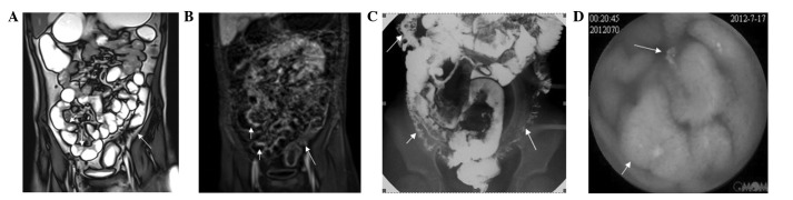
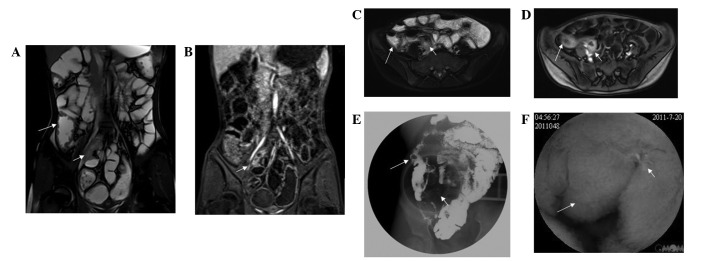
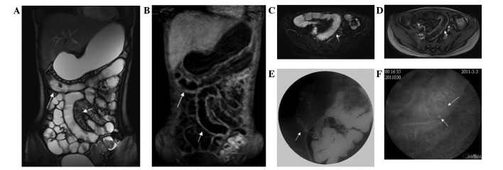
The aim of this study was to compare magnetic resonance enterography (MRE) findings with those of video capsule endoscopy (VCE) or conventional gastrointestinal radiography (CGR) in pediatric patients with small bowel Crohn's disease. A total of 55 cases of small bowel Crohn's disease that were diagnosed through clinical, laboratory, surgical and histopathological findings were reviewed. Prior to the examination, children suspected of having other types of diseases of the small intestinal were identified. The pulse sequences included coronal T2-true-fast imaging with steady-state precession (TrueFISP) images, navigation axial and coronal T1-weighted images, T2-weighted fat-suppressed images and coronal fat-suppressed three-dimensional gradient-echo images, immediately followed by contrast-enhanced axial and coronal T1-weighted fat-suppressed images. Findings from MRE were compared with those of VCE (n=39) and CGR (n=37). MRE results exhibited a number of features characteristic to small bowel Crohn's disease, including wall thickening, mesenteric fibrofatty changes and mesenteric vasculature changes. VCE, MRE and CGR demonstrated sensitivities of 94.6, 85.7 and 71.1% with specificities of 72.7, 70 and 40%; accuracies of 89.6, 82.2 and 61.1%; positive predictive values of 92.1, 90.9 and 59.6%; and negative predictive values of 80, 58.3 and 40%, respectively. VCE depicted mucosal pathologies missed by MRE in three patients. MRE revealed 83 extraenteric findings in 55 patients and CGR was able to show the dynamic evolution of the gastrointestinal function. MRE is a simple, safe, non-invasive and effective method for evaluating small bowel Crohn's disease. VCE allows visualization and readily characterizes subtle mucosal lesions missed by MRE, whereas MRE yields additional mural, perienteric and extraenteric information. However, oral barium CGR utilizes radiation, which is not suitable for repeated use in children.
本研究旨在比较磁共振小肠造影(MRE)与视频胶囊内镜(VCE)或传统胃肠造影(CGR)在小儿小肠克罗恩病患者中的检查结果。回顾了总共55例经临床、实验室、手术及组织病理学检查确诊的小肠克罗恩病病例。在检查前,先排除疑似患有其他类型小肠疾病的儿童。脉冲序列包括冠状位T2加权稳态进动快速成像(TrueFISP)图像、导航轴位和冠状位T1加权图像、T2加权脂肪抑制图像以及冠状位脂肪抑制三维梯度回波图像,随后立即进行对比增强轴位和冠状位T1加权脂肪抑制图像检查。将MRE的检查结果与VCE(n = 39)和CGR(n = 37)的结果进行比较。MRE结果显示出小肠克罗恩病的一些特征性表现,包括肠壁增厚、肠系膜纤维脂肪改变和肠系膜血管改变。VCE、MRE和CGR的敏感性分别为94.6%、85.7%和71.1%,特异性分别为72.7%、70%和40%;准确率分别为89.6%、82.2%和61.1%;阳性预测值分别为92.1%、90.9%和59.6%;阴性预测值分别为80%、58.3%和40%。VCE发现了3例MRE漏诊的黏膜病变。MRE在55例患者中发现了83处肠外病变,CGR则能够显示胃肠功能的动态演变。MRE是一种评估小肠克罗恩病的简单、安全、无创且有效的方法。VCE能够可视化并易于识别MRE漏诊的细微黏膜病变,而MRE可提供额外的肠壁、肠周和肠外信息。然而,口服钡剂CGR会产生辐射,不适用于儿童反复检查。