• 文献检索
  • 文档翻译
  • 深度研究
  • 学术资讯
  • Suppr Zotero 插件Zotero 插件
  • 邀请有礼
  • 套餐&价格
  • 历史记录
应用&插件
Suppr Zotero 插件Zotero 插件浏览器插件Mac 客户端Windows 客户端微信小程序
定价
高级版会员购买积分包购买API积分包
服务
文献检索文档翻译深度研究API 文档MCP 服务
关于我们
关于 Suppr公司介绍联系我们用户协议隐私条款
关注我们

Suppr 超能文献

核心技术专利:CN118964589B侵权必究
粤ICP备2023148730 号-1Suppr @ 2026

文献检索

告别复杂PubMed语法,用中文像聊天一样搜索,搜遍4000万医学文献。AI智能推荐,让科研检索更轻松。

立即免费搜索

文件翻译

保留排版,准确专业,支持PDF/Word/PPT等文件格式,支持 12+语言互译。

免费翻译文档

深度研究

AI帮你快速写综述,25分钟生成高质量综述,智能提取关键信息,辅助科研写作。

立即免费体验

定量分析抗原特异性细胞毒性过程中单个 CD8+ T 细胞的生物量变化。

Quantifying biomass changes of single CD8+ T cells during antigen specific cytotoxicity.

机构信息

Department of Pathology and Laboratory Medicine, University of California Los Angeles, Los Angeles, California, United States of America.

出版信息

PLoS One. 2013 Jul 23;8(7):e68916. doi: 10.1371/journal.pone.0068916. Print 2013.

DOI:10.1371/journal.pone.0068916
PMID:23935904
原文链接:https://pmc.ncbi.nlm.nih.gov/articles/PMC3720853/
Abstract

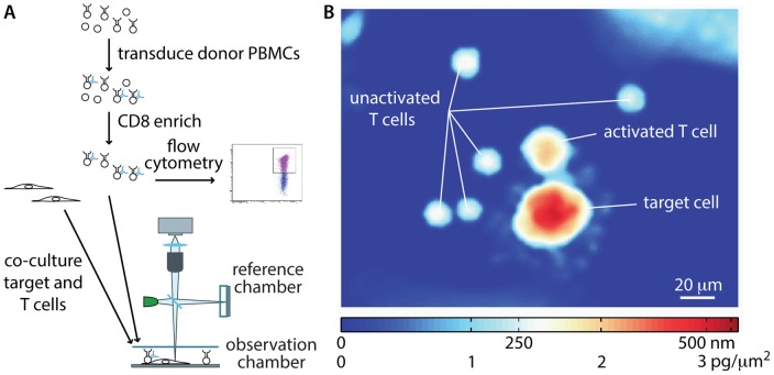
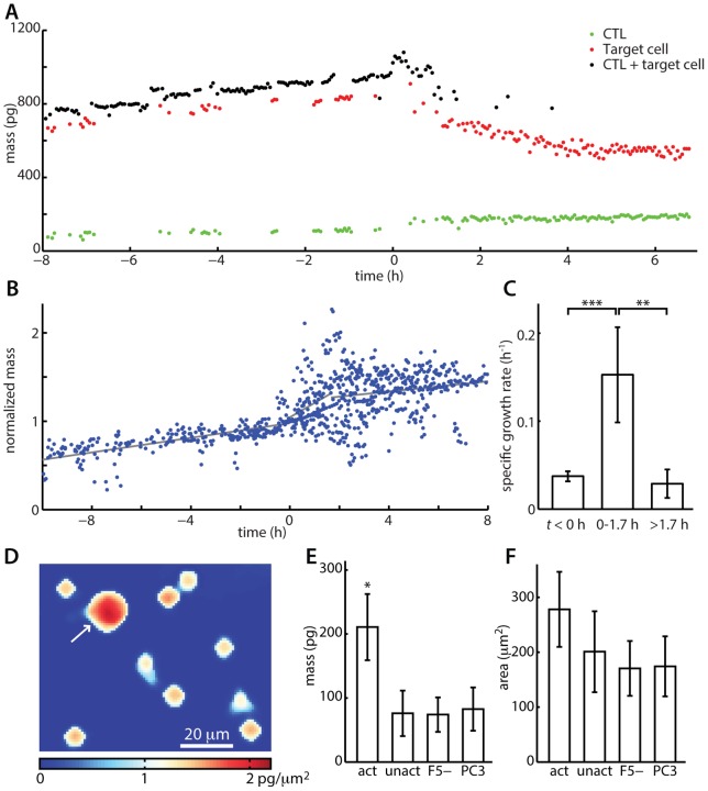
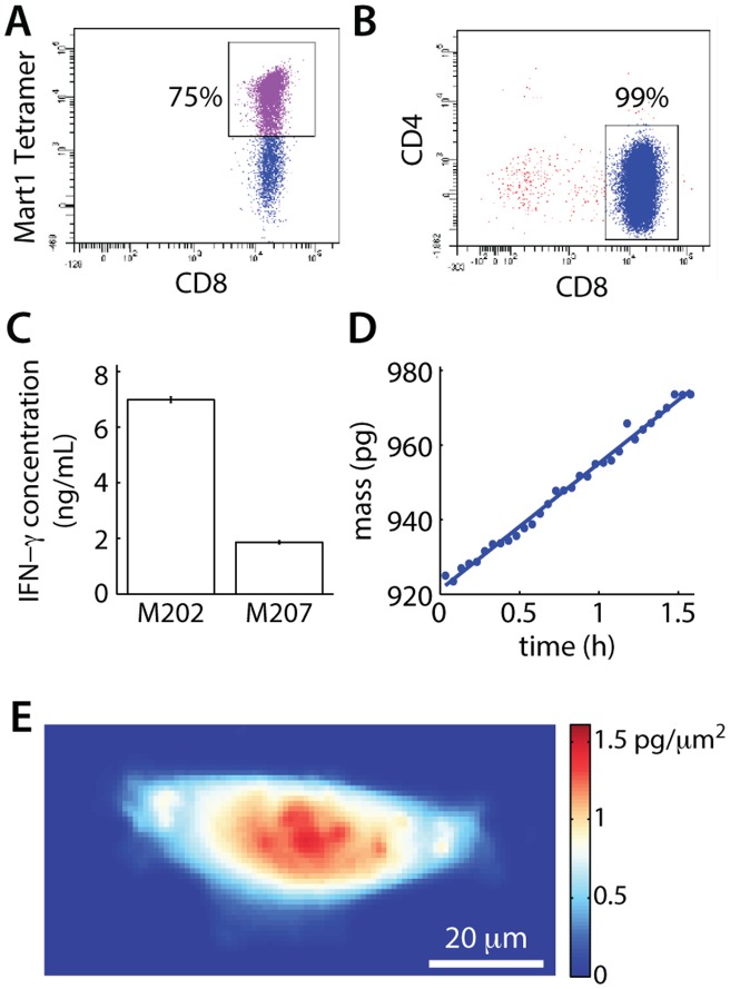
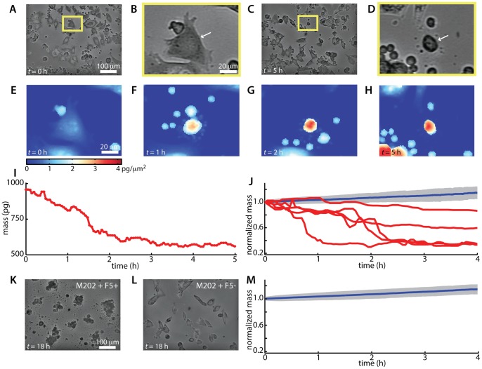
Existing approaches that quantify cytotoxic T cell responses rely on bulk or surrogate measurements which impede the direct identification of single activated T cells of interest. Single cell microscopy or flow cytometry methodologies typically rely on fluorescent labeling, which limits applicability to primary cells such as human derived T lymphocytes. Here, we introduce a quantitative method to track single T lymphocyte mediated cytotoxic events within a mixed population of cells using live cell interferometry (LCI), a label-free microscopy technique that maintains cell viability. LCI quantifies the mass distribution within individual cells by measuring the phase shift caused by the interaction of light with intracellular biomass. Using LCI, we imaged cytotoxic T cells killing cognate target cells. In addition to a characteristic target cell mass decrease of 20-60% over 1-4 h following attack by a T cell, there was a significant 4-fold increase in T cell mass accumulation rate at the start of the cytotoxic event and a 2-3 fold increase in T cell mass relative to the mass of unresponsive T cells. Direct, label-free measurement of CD8+ T and target cell mass changes provides a kinetic, quantitative assessment of T cell activation and a relatively rapid approach to identify specific, activated patient-derived T cells for applications in cancer immunotherapy.

摘要

现有的量化细胞毒性 T 细胞反应的方法依赖于批量或替代测量,这阻碍了对感兴趣的单个活化 T 细胞的直接识别。单细胞显微镜或流式细胞术方法通常依赖于荧光标记,这限制了其在原代细胞(如人类来源的 T 淋巴细胞)中的应用。在这里,我们引入了一种使用活细胞干涉测量(LCI)的定量方法,在混合细胞群中跟踪单个 T 淋巴细胞介导的细胞毒性事件,LCI 是一种无标记显微镜技术,可保持细胞活力。LCI 通过测量光与细胞内生物质相互作用引起的相移来量化单个细胞内的质量分布。使用 LCI,我们对细胞毒性 T 细胞杀伤同源靶细胞进行了成像。除了在 T 细胞攻击后 1-4 小时内靶细胞质量特征性减少 20-60%之外,在细胞毒性事件开始时 T 细胞的质量积累率显著增加了 4 倍,并且 T 细胞的质量相对于无反应性 T 细胞的质量增加了 2-3 倍。CD8+T 细胞和靶细胞质量变化的直接、无标记测量提供了 T 细胞活化的动力学、定量评估,并且是一种相对快速的方法,可以识别特定的、活化的患者来源的 T 细胞,用于癌症免疫治疗。

https://cdn.ncbi.nlm.nih.gov/pmc/blobs/d610/3720853/256a4675df44/pone.0068916.g004.jpg
https://cdn.ncbi.nlm.nih.gov/pmc/blobs/d610/3720853/1b44aa975526/pone.0068916.g001.jpg
https://cdn.ncbi.nlm.nih.gov/pmc/blobs/d610/3720853/07bba3e323a6/pone.0068916.g002.jpg
https://cdn.ncbi.nlm.nih.gov/pmc/blobs/d610/3720853/9e630e1b4fbb/pone.0068916.g003.jpg
https://cdn.ncbi.nlm.nih.gov/pmc/blobs/d610/3720853/256a4675df44/pone.0068916.g004.jpg

相似文献

1
Quantifying biomass changes of single CD8+ T cells during antigen specific cytotoxicity.定量分析抗原特异性细胞毒性过程中单个 CD8+ T 细胞的生物量变化。
PLoS One. 2013 Jul 23;8(7):e68916. doi: 10.1371/journal.pone.0068916. Print 2013.
2
Cytotoxic Activity and Memory T Cell Subset Distribution of -Stimulated CD8 T Cells Specific for HER2/neu Epitopes.针对 HER2/neu 表位的 - 刺激 CD8 T 细胞的细胞毒性活性和记忆 T 细胞亚群分布。
Front Immunol. 2019 May 9;10:1017. doi: 10.3389/fimmu.2019.01017. eCollection 2019.
3
A Method to Evaluate In Vivo CD8 T Cell Cytotoxicity in a Murine Model.一种评估小鼠模型体内 CD8 T 细胞细胞毒性的方法。
Methods Mol Biol. 2021;2183:549-558. doi: 10.1007/978-1-0716-0795-4_32.
4
Defining virus-specific CD8+ TCR repertoires for therapeutic regeneration of T cells against chronic hepatitis E.定义针对慢性戊型肝炎的 T 细胞治疗性再生的病毒特异性 CD8+ TCR 库。
J Hepatol. 2019 Oct;71(4):673-684. doi: 10.1016/j.jhep.2019.06.005. Epub 2019 Jun 14.
5
Usefulness of IL-21, IL-7, and IL-15 conditioned media for expansion of antigen-specific CD8+ T cells from healthy donor-PBMCs suitable for immunotherapy.IL-21、IL-7 和 IL-15 条件培养基在扩增健康供体-PBMC 中抗原特异性 CD8+T 细胞中的应用,适用于免疫治疗。
Cell Immunol. 2021 Feb;360:104257. doi: 10.1016/j.cellimm.2020.104257. Epub 2020 Dec 14.
6
Ex vivo measurement of the cytotoxic capacity of human primary antigen-specific CD8 T cells.体外测量人源原发性抗原特异性 CD8 T 细胞的细胞毒性能力。
J Immunol Methods. 2012 Jan 31;375(1-2):252-7. doi: 10.1016/j.jim.2011.09.008. Epub 2011 Oct 5.
7
A flow cytometry-based assay to assess minute frequencies of CD8+ T cells by their cytolytic function.基于流式细胞术的 assay,通过细胞溶解功能评估 CD8+ T 细胞的 minute 频率。
J Immunol Methods. 2010 Aug 31;360(1-2):56-65. doi: 10.1016/j.jim.2010.06.005. Epub 2010 Jun 15.
8
Activation of antigen-specific CD8 T cells results in minimal killing of bystander bacteria.抗原特异性CD8 T细胞的激活导致对旁观者细菌的杀伤作用极小。
J Immunol. 2003 Dec 1;171(11):6032-8. doi: 10.4049/jimmunol.171.11.6032.
9
[MHC tetramers: tracking specific immunity].[MHC四聚体:追踪特异性免疫]
Acta Med Croatica. 2003;57(4):255-9.
10
In vivo dynamics of anti-viral CD8 T cell responses to different epitopes. An evaluation of bystander activation in primary and secondary responses to viral infection.抗病毒CD8 T细胞对不同表位的体内动力学。对病毒感染初次和二次应答中旁观者激活的评估。
Adv Exp Med Biol. 1998;452:123-42. doi: 10.1007/978-1-4615-5355-7_14.

引用本文的文献

1
Tracking of lineage mass quantitative phase imaging and confinement in low refractive index microwells.对线形质量的追踪 定量相位成像及在低折射率微井中的限制。
Lab Chip. 2024 Sep 10;24(18):4440-4449. doi: 10.1039/d4lc00389f.
2
Engineering water exchange is a safe and effective method for magnetic resonance imaging in diverse cell types.工程水交换是一种用于多种细胞类型磁共振成像的安全有效方法。
J Biol Eng. 2024 Apr 22;18(1):30. doi: 10.1186/s13036-024-00424-5.
3
Deep learning-assisted smartphone-based quantitative microscopy for label-free peripheral blood smear analysis.

本文引用的文献

1
Rapid, massively parallel single-cell drug response measurements via live cell interferometry.通过活细胞干涉测量术实现快速、大规模平行的单细胞药物反应测量。
Biophys J. 2011 Sep 7;101(5):1025-31. doi: 10.1016/j.bpj.2011.07.022.
2
Optical measurement of cycle-dependent cell growth.细胞周期依赖性生长的光学测量
Proc Natl Acad Sci U S A. 2011 Aug 9;108(32):13124-9. doi: 10.1073/pnas.1100506108. Epub 2011 Jul 25.
3
A clinical microchip for evaluation of single immune cells reveals high functional heterogeneity in phenotypically similar T cells.
用于无标记外周血涂片分析的深度学习辅助智能手机定量显微镜技术。
Biomed Opt Express. 2024 Mar 27;15(4):2636-2651. doi: 10.1364/BOE.511384. eCollection 2024 Apr 1.
4
Engineering water exchange is a safe and effective method for magnetic resonance imaging in diverse cell types.工程水交换是一种用于多种细胞类型磁共振成像的安全有效方法。
bioRxiv. 2023 Nov 10:2023.11.07.566095. doi: 10.1101/2023.11.07.566095.
5
Quantitative Phase Imaging: Recent Advances and Expanding Potential in Biomedicine.定量相位成像:生物医学中的最新进展和扩展潜力。
ACS Nano. 2022 Aug 23;16(8):11516-11544. doi: 10.1021/acsnano.1c11507. Epub 2022 Aug 2.
6
Assessment of the electrical penetration of cell membranes using four-frequency impedance cytometry.使用四频阻抗细胞术评估细胞膜的电穿透情况。
Microsyst Nanoeng. 2022 Jun 24;8:68. doi: 10.1038/s41378-022-00405-y. eCollection 2022.
7
Transcriptional, Electrophysiological, and Metabolic Characterizations of hESC-Derived First and Second Heart Fields Demonstrate a Potential Role of TBX5 in Cardiomyocyte Maturation.人胚胎干细胞来源的第一和第二心脏区域的转录、电生理及代谢特征表明TBX5在心肌细胞成熟过程中的潜在作用。
Front Cell Dev Biol. 2021 Dec 17;9:787684. doi: 10.3389/fcell.2021.787684. eCollection 2021.
8
Cardiomyocytes disrupt pyrimidine biosynthesis in nonmyocytes to regulate heart repair.心肌细胞破坏非心肌细胞中的嘧啶生物合成以调节心脏修复。
J Clin Invest. 2022 Jan 18;132(2). doi: 10.1172/JCI149711.
9
Label-free automated neutropenia detection and grading using deep-ultraviolet microscopy.使用深紫外显微镜进行无标记自动中性粒细胞减少检测和分级。
Biomed Opt Express. 2021 Sep 9;12(10):6115-6128. doi: 10.1364/BOE.434465. eCollection 2021 Oct 1.
10
Rapid, label-free classification of tumor-reactive T cell killing with quantitative phase microscopy and machine learning.利用定量相位显微镜和机器学习快速、无标记地分类肿瘤反应性 T 细胞杀伤。
Sci Rep. 2021 Sep 30;11(1):19448. doi: 10.1038/s41598-021-98567-8.
一种用于评估单个免疫细胞的临床微芯片揭示了表型相似的 T 细胞中功能的高度异质性。
Nat Med. 2011 Jun;17(6):738-43. doi: 10.1038/nm.2375. Epub 2011 May 22.
4
T cell recognition of weak ligands: roles of signaling, receptor number, and affinity.T 细胞对弱配体的识别:信号转导、受体数量和亲和力的作用。
Immunol Res. 2011 May;50(1):39-48. doi: 10.1007/s12026-011-8204-3.
5
Optimizing optical flow cytometry for cell volume-based sorting and analysis.优化基于细胞体积的流式细胞术分选和分析。
PLoS One. 2011 Jan 20;6(1):e16053. doi: 10.1371/journal.pone.0016053.
6
Imaging techniques for assaying lymphocyte activation in action.用于检测淋巴细胞激活的成像技术。
Nat Rev Immunol. 2011 Jan;11(1):21-33. doi: 10.1038/nri2903.
7
The metabolic life and times of a T-cell.T 细胞的代谢生活及其时代
Immunol Rev. 2010 Jul;236:190-202. doi: 10.1111/j.1600-065X.2010.00911.x.
8
Differential sensitivity of melanoma cell lines with BRAFV600E mutation to the specific Raf inhibitor PLX4032.BRAFV600E 突变型黑色素瘤细胞系对特异性 Raf 抑制剂 PLX4032 的差异敏感性。
J Transl Med. 2010 Apr 20;8:39. doi: 10.1186/1479-5876-8-39.
9
The prioritization of cancer antigens: a national cancer institute pilot project for the acceleration of translational research.癌症抗原的优先级排序:美国国立癌症研究所加速转化研究的试点项目
Clin Cancer Res. 2009 Sep 1;15(17):5323-37. doi: 10.1158/1078-0432.CCR-09-0737.
10
Modular nucleic acid assembled p/MHC microarrays for multiplexed sorting of antigen-specific T cells.用于抗原特异性T细胞多重分选的模块化核酸组装的p/MHC微阵列。
J Am Chem Soc. 2009 Jul 22;131(28):9695-703. doi: 10.1021/ja9006707.