Hagey Laboratory, Department of Surgery, Stanford University School of Medicine, Stanford, California, USA.
PLoS One. 2013 Aug 1;8(8):e70240. doi: 10.1371/journal.pone.0070240. Print 2013.
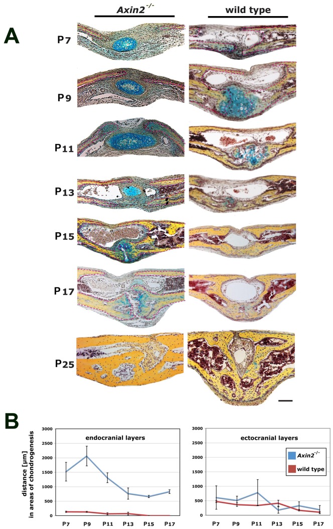
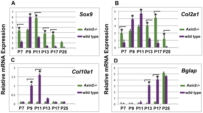
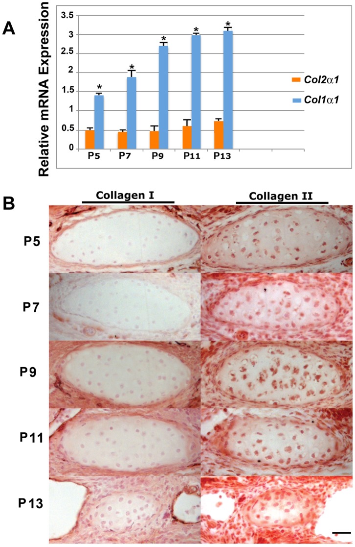
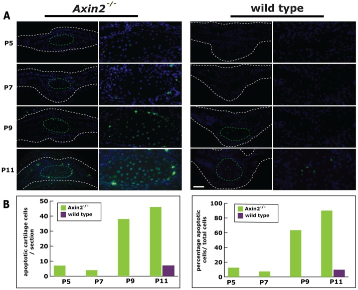
During the first month of life, the murine posterior-frontal suture (PF) of the cranial vault closes through endochondral ossification, while other sutures remain patent. These processes are tightly regulated by canonical Wnt signaling. Low levels of active canonical Wnt signaling enable endochondral ossification and therefore PF-suture closure, whereas constitutive activation of canonical Wnt causes PF-suture patency. We therefore sought to test this concept with a knockout mouse model. PF-sutures of Axin2(-/-) mice, which resemble a state of constantly activated canonical Wnt signaling, were investigated during the physiological time course of PF-suture closure and compared in detail with wild type littermates. Histological analysis revealed that the architecture in Axin2(-/-) PF-sutures was significantly altered in comparison to wild type. The distance between the endocranial layers was dramatically increased and suture closure was significantly delayed. Moreover, physiological endochondral ossification did not occur, rather an ectopic cartilage appeared between the endocranial and ectocranial bone layers at P7 which eventually involutes at P13. Quantitative PCR analysis showed the lack of Col10α1 upregulation in Axin2(-/-) PF-suture. Immunohistochemistry and gene expression analysis also revealed high levels of type II collagen as compared to type I collagen and absence of Mmp-9 in the cartilage of Axin2(-/-) PF-suture. Moreover, TUNEL staining showed a high percentage of apoptotic chondrocytes in Axin2(-/-) PF-sutures at P9 and P11 as compared to wild type. These data indicated that Axin2(-/-) PF-sutures lack physiological endochondral ossification, contain ectopic cartilage and display delayed suture closure.
在生命的第一个月,颅骨穹窿的鼠后额缝(PF)通过软骨内骨化闭合,而其他缝保持开放。这些过程受到经典 Wnt 信号通路的严格调控。低水平的活性经典 Wnt 信号通路可促进软骨内骨化,从而导致 PF 缝闭合,而经典 Wnt 的组成性激活导致 PF 缝开放。因此,我们试图用 knockout 小鼠模型来验证这一概念。在 PF 缝闭合的生理时间过程中,研究了 Axin2(-/-) 小鼠的 PF 缝,其类似于持续激活的经典 Wnt 信号通路的状态,并与野生型同窝仔鼠进行了详细比较。组织学分析表明,与野生型相比,Axin2(-/-) PF 缝的结构发生了显著改变。颅内层之间的距离显著增加,PF 缝闭合明显延迟。此外,生理软骨内骨化没有发生,而是在 P7 时在颅内层和外骨层之间出现异位软骨,最终在 P13 时退化。定量 PCR 分析表明,Axin2(-/-) PF 缝中 Col10α1 的上调缺乏。免疫组织化学和基因表达分析也显示,与 I 型胶原相比,Axin2(-/-) PF 缝中的 II 型胶原水平较高,且缺乏软骨中的 Mmp-9。此外,TUNEL 染色显示,与野生型相比,Axin2(-/-) PF 缝中的软骨细胞在 P9 和 P11 时凋亡率较高。这些数据表明,Axin2(-/-) PF 缝缺乏生理软骨内骨化,含有异位软骨,并显示出延迟的缝闭合。