• 文献检索
  • 文档翻译
  • 深度研究
  • 学术资讯
  • Suppr Zotero 插件Zotero 插件
  • 邀请有礼
  • 套餐&价格
  • 历史记录
应用&插件
Suppr Zotero 插件Zotero 插件浏览器插件Mac 客户端Windows 客户端微信小程序
定价
高级版会员购买积分包购买API积分包
服务
文献检索文档翻译深度研究API 文档MCP 服务
关于我们
关于 Suppr公司介绍联系我们用户协议隐私条款
关注我们

Suppr 超能文献

核心技术专利:CN118964589B侵权必究
粤ICP备2023148730 号-1Suppr @ 2026

文献检索

告别复杂PubMed语法,用中文像聊天一样搜索,搜遍4000万医学文献。AI智能推荐,让科研检索更轻松。

立即免费搜索

文件翻译

保留排版,准确专业,支持PDF/Word/PPT等文件格式,支持 12+语言互译。

免费翻译文档

深度研究

AI帮你快速写综述,25分钟生成高质量综述,智能提取关键信息,辅助科研写作。

立即免费体验

颅骨神经嵴细胞中骨形态发生蛋白信号增强通过软骨内成骨导致小鼠颅骨缝过早融合。

Augmentation of BMP Signaling in Cranial Neural Crest Cells Leads to Premature Cranial Sutures Fusion through Endochondral Ossification in Mice.

作者信息

Ueharu Hiroki, Pan Haichun, Liu Xia, Ishii Mamoru, Pongetti Jessica, Kulkarni Anshul K, Adegbenro Folasade E, Wurn Jaden, Maxson Robert E, Sun Hongchen, Komatsu Yoshihiro, Zhang Honghao, Yang Jingwen, Mishina Yuji

机构信息

Department of Biologic and Materials Sciences & Prosthodontics University Michigan School of Dentistry Ann Arbor Michigan USA.

Department of Oral Pathology Hospital of Stomatology, Jilin University Changchun China.

出版信息

JBMR Plus. 2023 Feb 23;7(4):e10716. doi: 10.1002/jbm4.10716. eCollection 2023 Apr.

DOI:10.1002/jbm4.10716
PMID:37065628
原文链接:https://pmc.ncbi.nlm.nih.gov/articles/PMC10097634/
Abstract

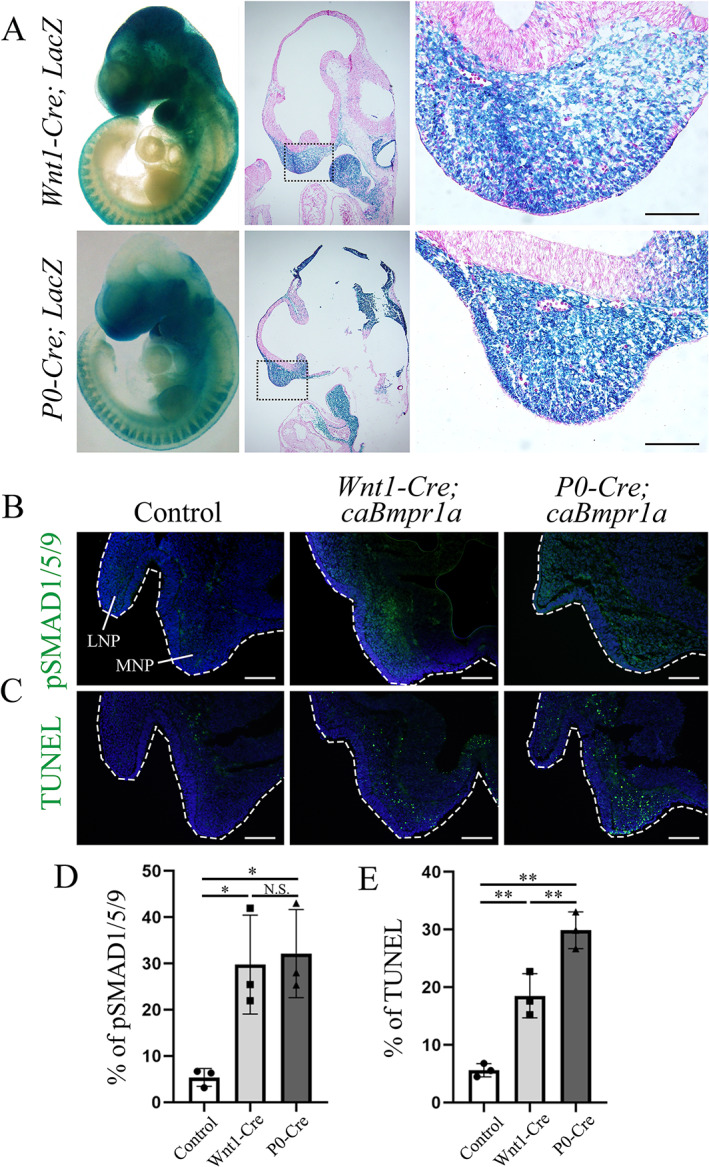
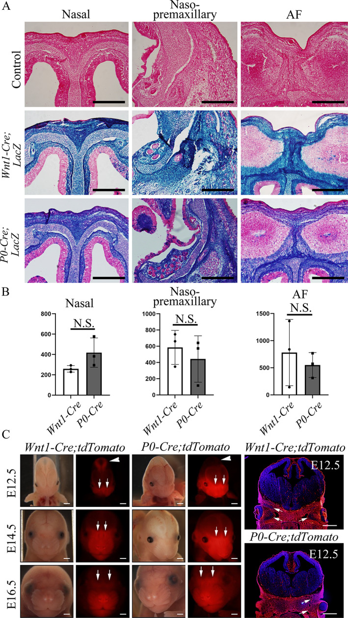
Craniosynostosis is a congenital anomaly characterized by the premature fusion of cranial sutures. Sutures are a critical connective tissue that regulates bone growth; their aberrant fusion results in abnormal shapes of the head and face. The molecular and cellular mechanisms have been investigated for a long time, but knowledge gaps remain between genetic mutations and mechanisms of pathogenesis for craniosynostosis. We previously demonstrated that the augmentation of bone morphogenetic protein (BMP) signaling through constitutively active BMP type 1A receptor (caBmpr1a) in neural crest cells (NCCs) caused the development of premature fusion of the anterior frontal suture, leading to craniosynostosis in mice. In this study, we demonstrated that ectopic cartilage forms in sutures prior to premature fusion in mice. The ectopic cartilage is subsequently replaced by bone nodules leading to premature fusion with similar but unique fusion patterns between two neural crest-specific transgenic Cre mouse lines, and mice, which coincides with patterns of premature fusion in each line. Histologic and molecular analyses suggest that endochondral ossification in the affected sutures. Both in vitro and in vivo observations suggest a greater chondrogenic capacity and reduced osteogenic capability of neural crest progenitor cells in mutant lines. These results suggest that the augmentation of BMP signaling alters the cell fate of cranial NCCs toward a chondrogenic lineage to prompt endochondral ossification to prematurely fuse cranial sutures. By comparing and mice at the stage of neural crest formation, we found more cell death of cranial NCCs in than mice at the developing facial primordia. These findings may provide a platform for understanding why mutations of broadly expressed genes result in the premature fusion of limited sutures. © 2022 The Authors. published by Wiley Periodicals LLC on behalf of American Society for Bone and Mineral Research.

摘要

颅缝早闭是一种先天性异常,其特征为颅骨缝过早融合。颅骨缝是调节骨骼生长的关键结缔组织;其异常融合会导致头面部形状异常。分子和细胞机制已被研究很长时间,但颅缝早闭的基因突变与发病机制之间仍存在知识空白。我们之前证明,通过在神经嵴细胞(NCCs)中组成型激活骨形态发生蛋白(BMP)1A型受体(caBmpr1a)来增强BMP信号传导,会导致前额缝过早融合,从而在小鼠中引发颅缝早闭。在本研究中,我们证明在小鼠过早融合之前,缝线中会形成异位软骨。随后,异位软骨被骨结节取代,导致在两种神经嵴特异性转基因Cre小鼠品系( 和 小鼠)之间出现类似但独特的融合模式的过早融合,这与每个品系中的过早融合模式一致。组织学和分子分析表明,受影响的缝线中存在软骨内成骨。体外和体内观察均表明,突变品系中的神经嵴祖细胞具有更大的软骨形成能力和更低的成骨能力。这些结果表明,BMP信号传导的增强会改变颅NCCs的细胞命运,使其向软骨形成谱系发展,从而促使软骨内成骨过早融合颅骨缝。通过比较神经嵴形成阶段的 和 小鼠,我们发现在发育中的面部原基处, 小鼠的颅NCCs细胞死亡比 小鼠更多。这些发现可能为理解为什么广泛表达的基因的突变会导致有限的缝线过早融合提供一个平台。© 2022作者。由Wiley Periodicals LLC代表美国骨与矿物质研究学会出版。

https://cdn.ncbi.nlm.nih.gov/pmc/blobs/8135/10097634/b790ba9dacef/JBM4-7-e10716-g001.jpg
https://cdn.ncbi.nlm.nih.gov/pmc/blobs/8135/10097634/6171328e88d2/JBM4-7-e10716-g002.jpg
https://cdn.ncbi.nlm.nih.gov/pmc/blobs/8135/10097634/bcd97e3142be/JBM4-7-e10716-g004.jpg
https://cdn.ncbi.nlm.nih.gov/pmc/blobs/8135/10097634/8164777a676b/JBM4-7-e10716-g005.jpg
https://cdn.ncbi.nlm.nih.gov/pmc/blobs/8135/10097634/10c46d2e81f4/JBM4-7-e10716-g006.jpg
https://cdn.ncbi.nlm.nih.gov/pmc/blobs/8135/10097634/208833bf636d/JBM4-7-e10716-g003.jpg
https://cdn.ncbi.nlm.nih.gov/pmc/blobs/8135/10097634/a43251ff1004/JBM4-7-e10716-g007.jpg
https://cdn.ncbi.nlm.nih.gov/pmc/blobs/8135/10097634/b790ba9dacef/JBM4-7-e10716-g001.jpg

相似文献

1
Augmentation of BMP Signaling in Cranial Neural Crest Cells Leads to Premature Cranial Sutures Fusion through Endochondral Ossification in Mice.颅骨神经嵴细胞中骨形态发生蛋白信号增强通过软骨内成骨导致小鼠颅骨缝过早融合。
JBMR Plus. 2023 Feb 23;7(4):e10716. doi: 10.1002/jbm4.10716. eCollection 2023 Apr.
2
Augmentation of bone morphogenetic protein signaling in cranial neural crest cells in mice deforms skull base due to premature fusion of intersphenoidal synchondrosis.在小鼠颅神经嵴细胞中增强骨形态发生蛋白信号会导致蝶鞍间软骨过早融合,从而使颅底变形。
Genesis. 2023 Mar;61(1-2):e23509. doi: 10.1002/dvg.23509. Epub 2023 Jan 9.
3
Augmentation of Smad-dependent BMP signaling in neural crest cells causes craniosynostosis in mice.在颅嵴细胞中增强 Smad 依赖性 BMP 信号会导致小鼠颅缝早闭。
J Bone Miner Res. 2013 Jun;28(6):1422-33. doi: 10.1002/jbmr.1857.
4
Rapamycin rescues BMP mediated midline craniosynostosis phenotype through reduction of mTOR signaling in a mouse model.在小鼠模型中,雷帕霉素通过降低mTOR信号传导挽救BMP介导的中线颅缝早闭表型。
Genesis. 2018 Jun;56(6-7):e23220. doi: 10.1002/dvg.23220.
5
Differential activation of canonical Wnt signaling determines cranial sutures fate: a novel mechanism for sagittal suture craniosynostosis.经典 Wnt 信号通路的差异激活决定颅缝命运:矢状缝颅缝早闭的新机制。
Dev Biol. 2010 Aug 15;344(2):922-40. doi: 10.1016/j.ydbio.2010.06.009. Epub 2010 Jun 12.
6
Pth1r in Neural Crest Cells Regulates Nasal Cartilage Differentiation.Pth1r 在神经嵴细胞中调节鼻软骨分化。
J Dent Res. 2024 Mar;103(3):308-317. doi: 10.1177/00220345231221954. Epub 2024 Jan 17.
7
Specific and spatial labeling of P0-Cre versus Wnt1-Cre in cranial neural crest in early mouse embryos.早期小鼠胚胎中颅神经嵴中P0-Cre与Wnt1-Cre的特异性和空间标记。
Genesis. 2017 Jun;55(6). doi: 10.1002/dvg.23034. Epub 2017 Apr 18.
8
Identification of progenitor cells that contribute to heterotopic skeletogenesis.对促成异位骨骼形成的祖细胞的鉴定。
J Bone Joint Surg Am. 2009 Mar 1;91(3):652-63. doi: 10.2106/JBJS.H.01177.
9
Cranial Neural Crest Cells and Their Role in the Pathogenesis of Craniofacial Anomalies and Coronal Craniosynostosis.颅神经嵴细胞及其在颅面畸形和冠状缝早闭发病机制中的作用。
J Dev Biol. 2020 Sep 9;8(3):18. doi: 10.3390/jdb8030018.
10
BmpR1A is a major type 1 BMP receptor for BMP-Smad signaling during skull development.BmpR1A是颅骨发育过程中BMP-Smad信号传导的主要I型BMP受体。
Dev Biol. 2017 Sep 1;429(1):260-270. doi: 10.1016/j.ydbio.2017.06.020. Epub 2017 Jun 19.

引用本文的文献

1
The tectum transversum(TTR) maintains patency of the developing coronal suture.中脑顶盖横部(TTR)维持发育中冠状缝的通畅。
Res Sq. 2025 Aug 5:rs.3.rs-7216722. doi: 10.21203/rs.3.rs-7216722/v1.
2
Augmented Bone Morphogenetic Protein Signaling During TMJ Development Alters Morphology in a Timepoint-Dependent Manner.颞下颌关节发育过程中骨形态发生蛋白信号增强以时间点依赖的方式改变形态。
Int J Mol Sci. 2025 Feb 15;26(4):1655. doi: 10.3390/ijms26041655.
3
Polarity and migration of cranial and cardiac neural crest cells: underlying molecular mechanisms and disease implications.

本文引用的文献

1
Scaffold Pore Curvature Influences ΜSC Fate through Differential Cellular Organization and YAP/TAZ Activity.支架孔曲率通过细胞的不同组织和 YAP/TAZ 活性影响间充质干细胞命运。
Int J Mol Sci. 2022 Apr 19;23(9):4499. doi: 10.3390/ijms23094499.
2
Quantification of three-dimensional morphology of craniofacial mineralized tissue defects in Tgfbr2/Osx-Cre mice.Tgfbr2/Osx-Cre小鼠颅面矿化组织缺损的三维形态定量分析
Oral Sci Int. 2021 Sep;18(3):193-202. doi: 10.1002/osi2.1099. Epub 2021 Jan 8.
3
Riding the crest to get a head: neural crest evolution in vertebrates.
颅面和心脏神经嵴细胞的极性与迁移:潜在分子机制及疾病影响
Front Cell Dev Biol. 2025 Jan 6;12:1457506. doi: 10.3389/fcell.2024.1457506. eCollection 2024.
4
Timing and Graded BMP Signalling Determines Fate of Neural Crest and Ectodermal Placode Derivatives from Pluripotent Stem Cells.时间和分级BMP信号决定多能干细胞来源的神经嵴和外胚层基板衍生物的命运。
Biomedicines. 2024 Oct 4;12(10):2262. doi: 10.3390/biomedicines12102262.
5
Transforming growth factor beta signaling and craniofacial development: modeling human diseases in zebrafish.转化生长因子β信号传导与颅面发育:在斑马鱼中模拟人类疾病
Front Cell Dev Biol. 2024 Feb 7;12:1338070. doi: 10.3389/fcell.2024.1338070. eCollection 2024.
6
BMP signaling during craniofacial development: new insights into pathological mechanisms leading to craniofacial anomalies.颅面发育过程中的骨形态发生蛋白信号传导:对导致颅面异常的病理机制的新见解。
Front Physiol. 2023 May 18;14:1170511. doi: 10.3389/fphys.2023.1170511. eCollection 2023.
乘风破浪迎头而上:脊椎动物神经嵴的演化。
Nat Rev Neurosci. 2021 Oct;22(10):616-626. doi: 10.1038/s41583-021-00503-2. Epub 2021 Sep 1.
4
BMPR1A maintains skeletal stem cell properties in craniofacial development and craniosynostosis.BMPR1A 维持颅面发育和颅缝早闭过程中的颅面干/基质细胞特性。
Sci Transl Med. 2021 Mar 3;13(583). doi: 10.1126/scitranslmed.abb4416.
5
Augmented BMP signaling commits cranial neural crest cells to a chondrogenic fate by suppressing autophagic β-catenin degradation.增强的 BMP 信号通过抑制自噬 β-连环蛋白降解来使颅神经嵴细胞向软骨生成命运分化。
Sci Signal. 2021 Jan 12;14(665):eaaz9368. doi: 10.1126/scisignal.aaz9368.
6
Controversy of physiological vs. pharmacological effects of BMP signaling: Constitutive activation of BMP type IA receptor-dependent signaling in osteoblast lineage enhances bone formation and resorption, not affecting net bone mass.骨形态发生蛋白(BMP)信号的生理与药理作用之争:成骨细胞系中 BMP Ⅰ型受体依赖性信号的组成性激活增强了骨形成和吸收,但不影响净骨量。
Bone. 2020 Sep;138:115513. doi: 10.1016/j.bone.2020.115513. Epub 2020 Jun 27.
7
A review of the management of single-suture craniosynostosis, past, present, and future.单缝颅缝早闭的治疗回顾:过去、现在与未来
J Neurosurg Pediatr. 2019 Dec 1;24(6):622-631. doi: 10.3171/2019.7.PEDS18585.
8
A Ciliary Protein EVC2/LIMBIN Plays a Critical Role in the Skull Base for Mid-Facial Development.一种纤毛蛋白EVC2/LIMBIN在颅底中部面部发育中起关键作用。
Front Physiol. 2018 Oct 25;9:1484. doi: 10.3389/fphys.2018.01484. eCollection 2018.
9
Rapamycin rescues BMP mediated midline craniosynostosis phenotype through reduction of mTOR signaling in a mouse model.在小鼠模型中,雷帕霉素通过降低mTOR信号传导挽救BMP介导的中线颅缝早闭表型。
Genesis. 2018 Jun;56(6-7):e23220. doi: 10.1002/dvg.23220.
10
Transcriptional network systems in cartilage development and disease.软骨发育与疾病中的转录网络系统
Histochem Cell Biol. 2018 Apr;149(4):353-363. doi: 10.1007/s00418-017-1628-7. Epub 2018 Jan 8.