• 文献检索
  • 文档翻译
  • 深度研究
  • 学术资讯
  • Suppr Zotero 插件Zotero 插件
  • 邀请有礼
  • 套餐&价格
  • 历史记录
应用&插件
Suppr Zotero 插件Zotero 插件浏览器插件Mac 客户端Windows 客户端微信小程序
定价
高级版会员购买积分包购买API积分包
服务
文献检索文档翻译深度研究API 文档MCP 服务
关于我们
关于 Suppr公司介绍联系我们用户协议隐私条款
关注我们

Suppr 超能文献

核心技术专利:CN118964589B侵权必究
粤ICP备2023148730 号-1Suppr @ 2026

文献检索

告别复杂PubMed语法,用中文像聊天一样搜索,搜遍4000万医学文献。AI智能推荐,让科研检索更轻松。

立即免费搜索

文件翻译

保留排版,准确专业,支持PDF/Word/PPT等文件格式,支持 12+语言互译。

免费翻译文档

深度研究

AI帮你快速写综述,25分钟生成高质量综述,智能提取关键信息,辅助科研写作。

立即免费体验

PET/MRI成像在高危肉瘤中的应用:初步发现及临床问题解决

PET/MRI Imaging in High-Risk Sarcoma: First Findings and Solving Clinical Problems.

作者信息

Schuler Markus K, Richter Stephan, Beuthien-Baumann Bettina, Platzek Ivan, Kotzerke Jörg, van den Hoff Jörg, Ehninger Gerhard, Reichardt Peter

机构信息

Department of Internal Medicine I, University Hospital Carl Gustav Carus, Technical University at Dresden, Fetscherstraße 74, 01307 Dresden, Germany.

出版信息

Case Rep Oncol Med. 2013;2013:793927. doi: 10.1155/2013/793927. Epub 2013 Jul 1.

DOI:10.1155/2013/793927
PMID:23936700
原文链接:https://pmc.ncbi.nlm.nih.gov/articles/PMC3713328/
Abstract

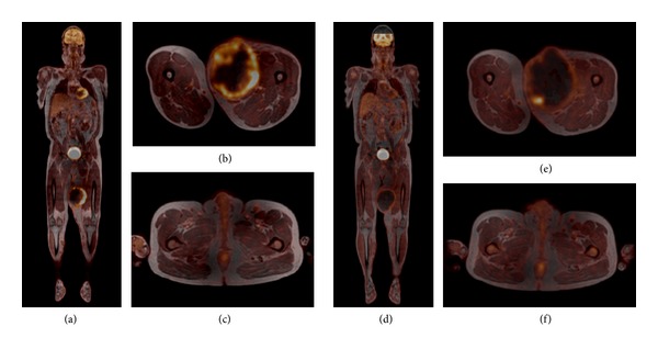
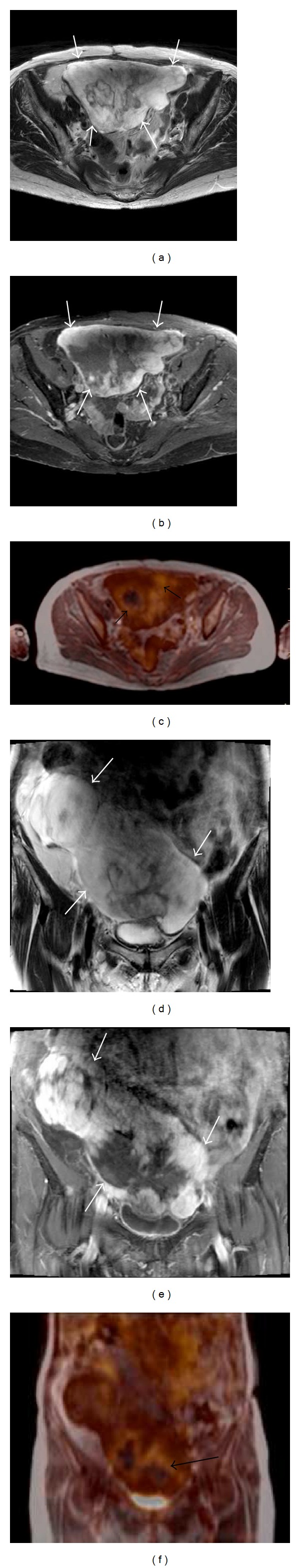
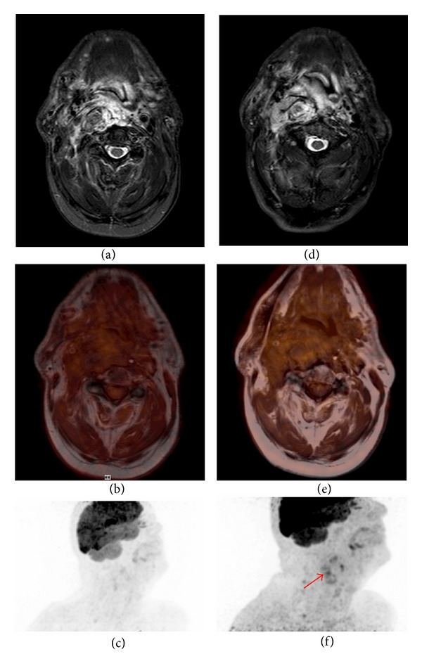
Simultaneous positron emission tomography (PET) and magnetic resonance imaging (MRI) is a new whole-body hybrid PET/MR imaging technique that combines metabolic and cross-sectional diagnostic imaging. Since the use of MRI in imaging of soft-tissue sarcoma is extremely beneficial, investigation of the combined PET/MRI is of great interest. In this paper, we present three cases and first data. Combined PET/MRI technique can support the process of clinical decision-making and give answers to some meaningful questions when treating patients with STS. Therefore, the combined modality of simultaneous PET/MRI offers new pieces to the puzzle of sarcoma treatment.

摘要

同时正电子发射断层扫描(PET)和磁共振成像(MRI)是一种新型的全身混合PET/MR成像技术,它结合了代谢和断层诊断成像。由于MRI在软组织肉瘤成像中的应用极其有益,因此对PET/MRI联合成像的研究备受关注。在本文中,我们展示了三个病例及初步数据。PET/MRI联合技术能够支持临床决策过程,并在治疗软组织肉瘤患者时回答一些有意义的问题。因此,同时进行PET/MRI的联合模式为肉瘤治疗难题提供了新的线索。

https://cdn.ncbi.nlm.nih.gov/pmc/blobs/dc47/3713328/b1fe2b964702/CRIM.ONCMED2013-793927.003.jpg
https://cdn.ncbi.nlm.nih.gov/pmc/blobs/dc47/3713328/4969b69964cf/CRIM.ONCMED2013-793927.001.jpg
https://cdn.ncbi.nlm.nih.gov/pmc/blobs/dc47/3713328/da348ba061fa/CRIM.ONCMED2013-793927.002.jpg
https://cdn.ncbi.nlm.nih.gov/pmc/blobs/dc47/3713328/b1fe2b964702/CRIM.ONCMED2013-793927.003.jpg
https://cdn.ncbi.nlm.nih.gov/pmc/blobs/dc47/3713328/4969b69964cf/CRIM.ONCMED2013-793927.001.jpg
https://cdn.ncbi.nlm.nih.gov/pmc/blobs/dc47/3713328/da348ba061fa/CRIM.ONCMED2013-793927.002.jpg
https://cdn.ncbi.nlm.nih.gov/pmc/blobs/dc47/3713328/b1fe2b964702/CRIM.ONCMED2013-793927.003.jpg

相似文献

1
PET/MRI Imaging in High-Risk Sarcoma: First Findings and Solving Clinical Problems.PET/MRI成像在高危肉瘤中的应用:初步发现及临床问题解决
Case Rep Oncol Med. 2013;2013:793927. doi: 10.1155/2013/793927. Epub 2013 Jul 1.
2
The impact of F-FDG PET on initial staging and therapy planning of pediatric soft-tissue sarcoma patients.氟代脱氧葡萄糖正电子发射断层扫描对儿科软组织肉瘤患者初始分期和治疗计划的影响。
Pediatr Radiol. 2020 Feb;50(2):252-260. doi: 10.1007/s00247-019-04530-1. Epub 2019 Oct 18.
3
Synergistic role of simultaneous PET/MRI-MRS in soft tissue sarcoma metabolism imaging.正电子发射断层扫描/磁共振成像-磁共振波谱同时成像在软组织肉瘤代谢成像中的协同作用。
Magn Reson Imaging. 2016 Apr;34(3):276-9. doi: 10.1016/j.mri.2015.10.027. Epub 2015 Oct 31.
4
Magnetic resonance imaging and positron emission tomography-computed tomography evaluation of soft tissue sarcoma with surgical and histopathological correlation.软组织肉瘤的磁共振成像及正电子发射断层扫描-计算机断层扫描评估与手术及组织病理学相关性研究
Indian J Nucl Med. 2012 Oct;27(4):213-20. doi: 10.4103/0972-3919.115390.
5
Evaluation of diagnostic performance of whole-body simultaneous PET/MRI in pediatric lymphoma.儿童淋巴瘤全身同步PET/MRI诊断性能评估
Pediatr Radiol. 2016 Aug;46(9):1258-68. doi: 10.1007/s00247-016-3601-3. Epub 2016 Mar 22.
6
Fluorodeoxyglucose positron-emission tomography-magnetic resonance hybrid imaging: An emerging tool for staging of cancer of the uterine cervix.氟脱氧葡萄糖正电子发射断层扫描-磁共振混合成像:一种用于宫颈癌分期的新兴工具。
World J Nucl Med. 2020 Aug 22;20(2):150-155. doi: 10.4103/wjnm.WJNM_53_20. eCollection 2021 Apr-Jun.
7
Competitive advantage of PET/MRI.正电子发射断层扫描/磁共振成像的竞争优势。
Eur J Radiol. 2014 Jan;83(1):84-94. doi: 10.1016/j.ejrad.2013.05.028. Epub 2013 Jun 18.
8
Comparison of MRI and PET-CT in detecting the loco-regional recurrence of soft tissue sarcomas during surveillance.MRI与PET-CT在监测软组织肉瘤局部区域复发中的比较。
Skeletal Radiol. 2016 Oct;45(10):1375-84. doi: 10.1007/s00256-016-2440-5. Epub 2016 Aug 3.
9
Hybrid PET/MR imaging in two sarcoma patients - clinical benefits and implications for future trials.两名肉瘤患者的PET/MR混合成像——临床益处及对未来试验的启示
Int J Clin Exp Med. 2014 Mar 15;7(3):640-8. eCollection 2014.
10
Future Perspective of the Application of Positron Emission Tomography-Computed Tomography-MR Imaging in Musculoskeletal Disorders.正电子发射断层扫描-计算机断层扫描-磁共振成像在肌肉骨骼疾病中的应用前景
PET Clin. 2019 Jan;14(1):183-191. doi: 10.1016/j.cpet.2018.08.012. Epub 2018 Oct 24.

引用本文的文献

1
Applications of PET-MRI in musculoskeletal disease.正电子发射断层扫描-磁共振成像在肌肉骨骼疾病中的应用。
J Magn Reson Imaging. 2018 Jul;48(1):27-47. doi: 10.1002/jmri.26183.
2
Integrated 18F-FDG PET/MRI compared to MRI alone for identification of local recurrences of soft tissue sarcomas: a comparison trial.18F-FDG PET/MRI 与单独 MRI 对比用于软组织肉瘤局部复发的识别:一项对比试验。
Eur J Nucl Med Mol Imaging. 2017 Oct;44(11):1823-1831. doi: 10.1007/s00259-017-3736-y. Epub 2017 May 31.
3
MR-PET of the body: Early experience and insights.

本文引用的文献

1
Oncologic PET/MRI, part 2: bone tumors, soft-tissue tumors, melanoma, and lymphoma.肿瘤 PET/MRI 成像,第 2 部分:骨肿瘤、软组织肿瘤、黑色素瘤和淋巴瘤。
J Nucl Med. 2012 Aug;53(8):1244-52. doi: 10.2967/jnumed.112.109306. Epub 2012 Jul 10.
2
18F-FDG-PET/CT Imaging as an early survival predictor in patients with primary high-grade soft tissue sarcomas undergoing neoadjuvant therapy.18F-FDG-PET/CT 成像作为新辅助治疗后原发性高级别软组织肉瘤患者早期生存预测因子。
Clin Cancer Res. 2012 Apr 1;18(7):2024-31. doi: 10.1158/1078-0432.CCR-11-2139. Epub 2012 Feb 14.
3
3'-deoxy-3'-[18F]fluorothymidine positron emission tomography for response assessment in soft tissue sarcoma: a pilot study to correlate imaging findings with tissue thymidine kinase 1 and Ki-67 activity and histopathologic response.
身体的磁共振成像-正电子发射断层显像:早期经验与见解。
Eur J Radiol Open. 2014 Sep 16;1:28-39. doi: 10.1016/j.ejro.2014.09.001. eCollection 2014.
4
Integrated whole body MR/PET: where are we?全身MR/PET一体化成像:我们目前的进展如何?
Korean J Radiol. 2015 Jan-Feb;16(1):32-49. doi: 10.3348/kjr.2015.16.1.32. Epub 2015 Jan 9.
3'-去氧-3'-[18F]氟代胸腺嘧啶正电子发射断层扫描用于软组织肉瘤反应评估:一项与组织胸苷激酶 1 和 Ki-67 活性及组织病理学反应相关的成像发现的初步研究。
Cancer. 2012 Jun 15;118(12):3135-44. doi: 10.1002/cncr.26630. Epub 2011 Oct 21.
4
Contribution of magnetic resonance imaging in the diagnosis of talus skip metastases of Ewing's sarcoma of the calcaneus in a child: a case report.磁共振成像在诊断儿童跟骨尤因肉瘤距骨跳跃转移中的作用:一例报告
J Med Case Rep. 2011 Sep 12;5:451. doi: 10.1186/1752-1947-5-451.
5
PET/CT allows stratification of responders to neoadjuvant chemotherapy for high-grade sarcoma: a prospective study.PET/CT 可对高级别肉瘤新辅助化疗的应答者进行分层:一项前瞻性研究。
Clin Nucl Med. 2011 Jul;36(7):526-32. doi: 10.1097/RLU.0b013e3182175856.
6
Initial McGill experience with fluorodeoxyglucose pet/ct staging of soft-tissue sarcoma.软组织肉瘤氟脱氧葡萄糖 pet/ct 分期的初步麦吉尔经验。
Curr Oncol. 2010 Nov;17(6):18-22. doi: 10.3747/co.v17i6.538.
7
Soft tissue sarcomas: ESMO Clinical Practice Guidelines for diagnosis, treatment and follow-up.软组织肉瘤:ESMO诊断、治疗及随访临床实践指南
Ann Oncol. 2010 May;21 Suppl 5:v198-203. doi: 10.1093/annonc/mdq209.
8
Effect of adjuvant chemotherapy on survival in FNCLCC grade 3 soft tissue sarcomas: a multivariate analysis of the French Sarcoma Group Database.辅助化疗对 FNCLCC 分级 3 软组织肉瘤患者生存的影响:法国肉瘤研究组数据库的多变量分析。
Ann Oncol. 2010 Dec;21(12):2436-2441. doi: 10.1093/annonc/mdq238. Epub 2010 May 3.
9
Adjuvant chemotherapy in localized soft tissue sarcomas: still not proven.局部软组织肉瘤的辅助化疗:仍未得到证实。
Oncologist. 2009 Oct;14(10):1013-20. doi: 10.1634/theoncologist.2009-0126. Epub 2009 Oct 6.
10
Should patients with high-risk soft tissue sarcoma receive adjuvant chemotherapy?高危软组织肉瘤患者应该接受辅助化疗吗?
Oncologist. 2009 Oct;14(10):1003-12. doi: 10.1634/theoncologist.2009-0007. Epub 2009 Oct 6.