Nguyen John T, Lin Samuel J, Tobias Adam M, Gioux Sylvain, Mazhar Amaan, Cuccia David J, Ashitate Yoshitomo, Stockdale Alan, Oketokoun Rafiou, Durr Nicholas J, Moffitt Lorissa A, Durkin Anthony J, Tromberg Bruce J, Frangioni John V, Lee Bernard T
Division of Plastic and Reconstructive Surgery, Department of Surgery, Department of Medicine, Beth Israel Deaconess Medical Center, Boston, MA, USA.
Ann Plast Surg. 2013 Sep;71(3):308-15. doi: 10.1097/SAP.0b013e31828b02fb.
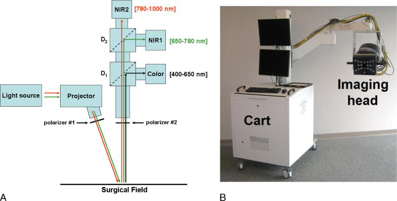
Although various methods exist for monitoring flaps during reconstructive surgery, surgeons primarily rely on assessment of clinical judgment. Early detection of vascular complications improves rate of flap salvage. Spatial frequency domain imaging (SFDI) is a promising new technology that provides oxygenation images over a large field of view. The goal of this clinical pilot study is to use SFDI in perforator flap breast reconstruction.
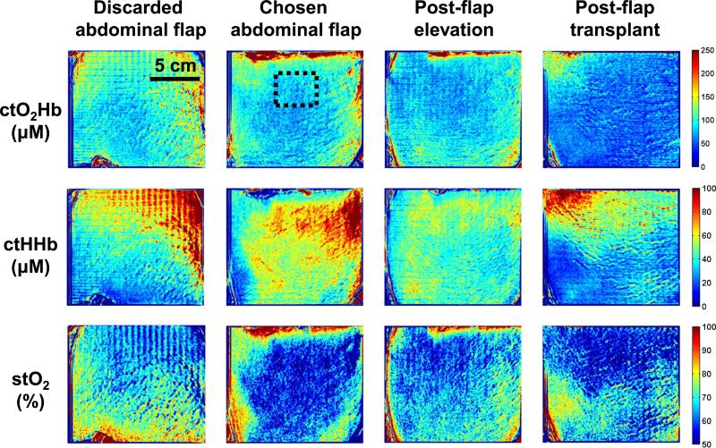
Three women undergoing unilateral breast reconstruction after mastectomy were enrolled for our study. The SFDI system was deployed in the operating room, and images acquired over the course of the operation. Time points included images of each hemiabdominal skin flap before elevation, the selected flap after perforator dissection, and after microsurgical transfer.
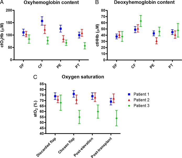
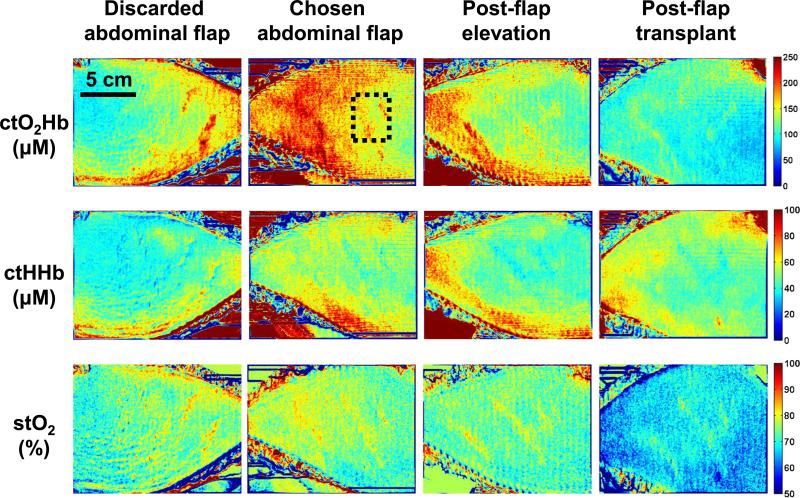
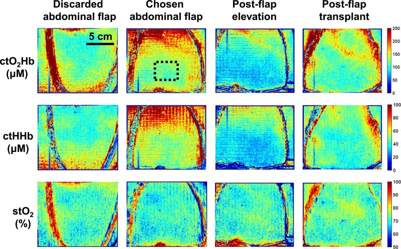
Spatial frequency domain imaging was able to measure tissue oxyhemoglobin concentration (ctO2Hb), tissue deoxyhemoglobin concentration, and tissue oxygen saturation (stO2). Images were created for each metric to monitor flap status and the results quantified throughout the various time points of the procedure. For 2 of 3 patients, the chosen flap had a higher ctO2Hb and stO2. For 1 patient, the chosen flap had lower ctO2Hb and stO2. There were no perfusion deficits observed based on SFDI and clinical follow-up.
The results of our initial human pilot study suggest that SFDI has the potential to provide intraoperative oxygenation images in real-time during surgery. With the use of this technology, surgeons can obtain tissue oxygenation and hemoglobin concentration maps to assist in intraoperative planning; this can potentially prevent complications and improve clinical outcome.
尽管在重建手术中存在多种监测皮瓣的方法,但外科医生主要依靠临床判断进行评估。早期发现血管并发症可提高皮瓣挽救率。空间频域成像(SFDI)是一项有前景的新技术,可在大视野范围内提供氧合图像。本临床初步研究的目的是在穿支皮瓣乳房重建中使用SFDI。
三名接受乳房切除术后单侧乳房重建的女性纳入本研究。SFDI系统部署在手术室,并在手术过程中采集图像。时间点包括每个半腹皮瓣掀起前、穿支解剖后所选皮瓣以及显微外科转移后的图像。
空间频域成像能够测量组织氧合血红蛋白浓度(ctO2Hb)、组织脱氧血红蛋白浓度和组织氧饱和度(stO2)。为每个指标创建图像以监测皮瓣状态,并在手术的各个时间点对结果进行量化。3例患者中有2例,所选皮瓣的ctO2Hb和stO2较高。1例患者所选皮瓣的ctO2Hb和stO2较低。基于SFDI和临床随访未观察到灌注不足。
我们最初的人体初步研究结果表明,SFDI有可能在手术期间实时提供术中氧合图像。通过使用这项技术,外科医生可以获得组织氧合和血红蛋白浓度图以协助术中规划;这有可能预防并发症并改善临床结果。