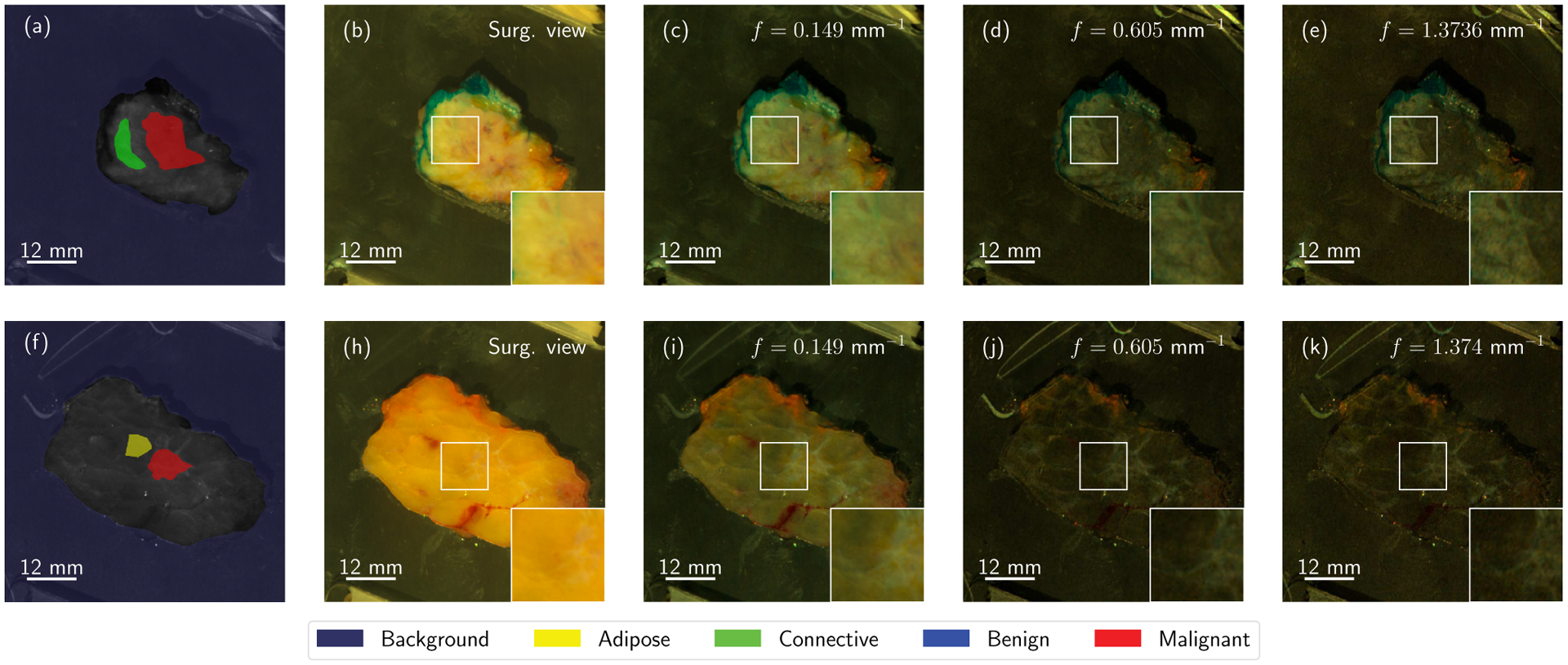
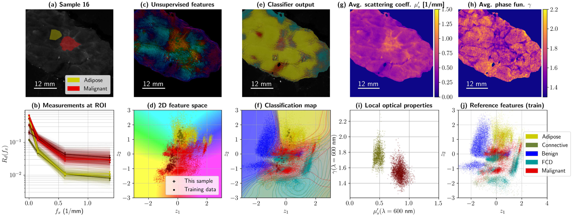
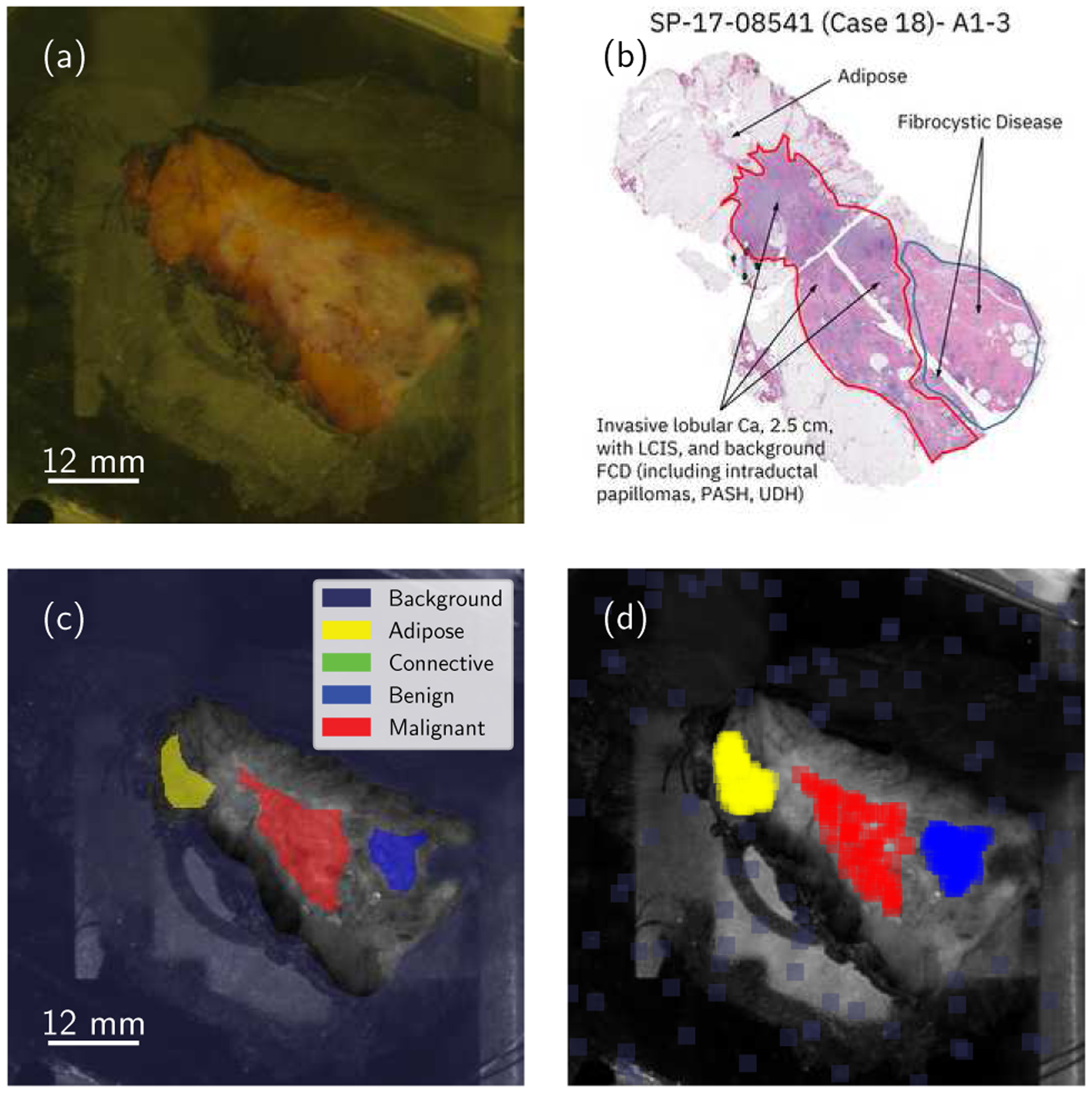
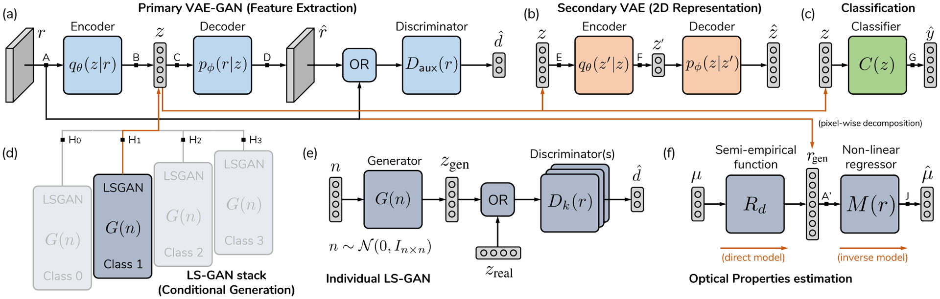
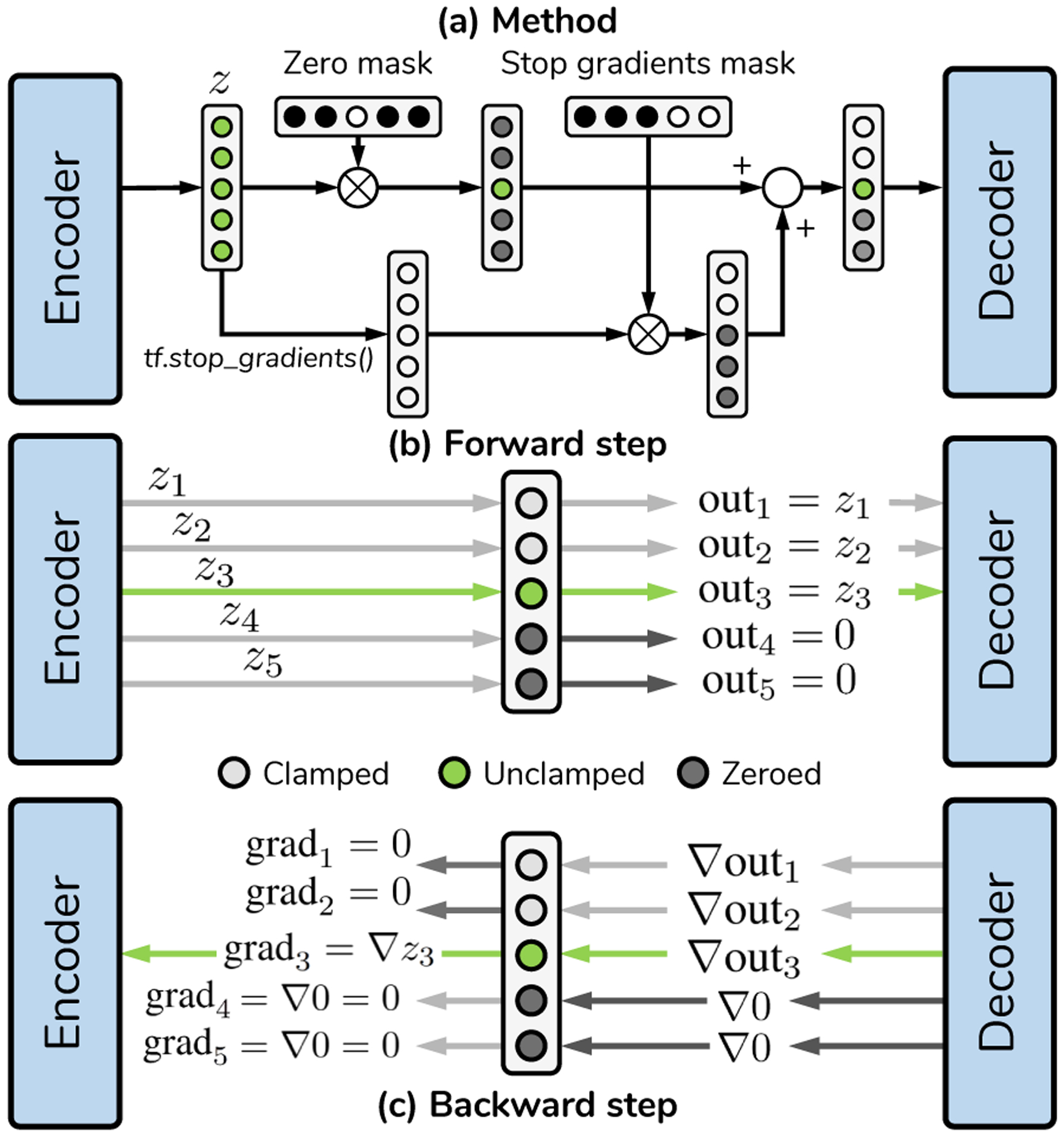
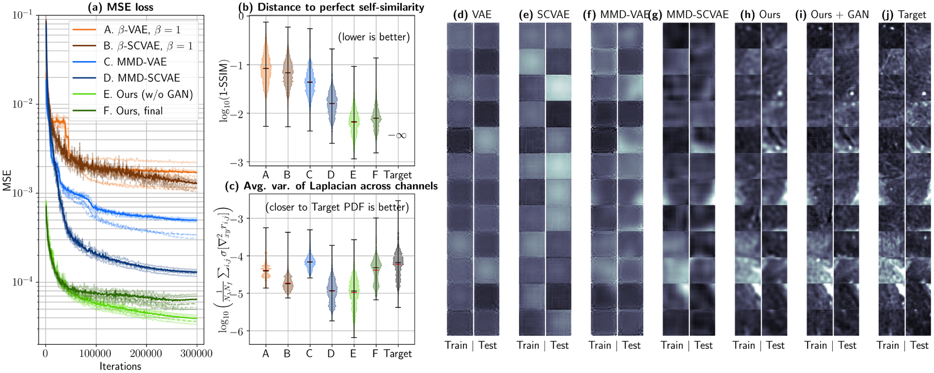
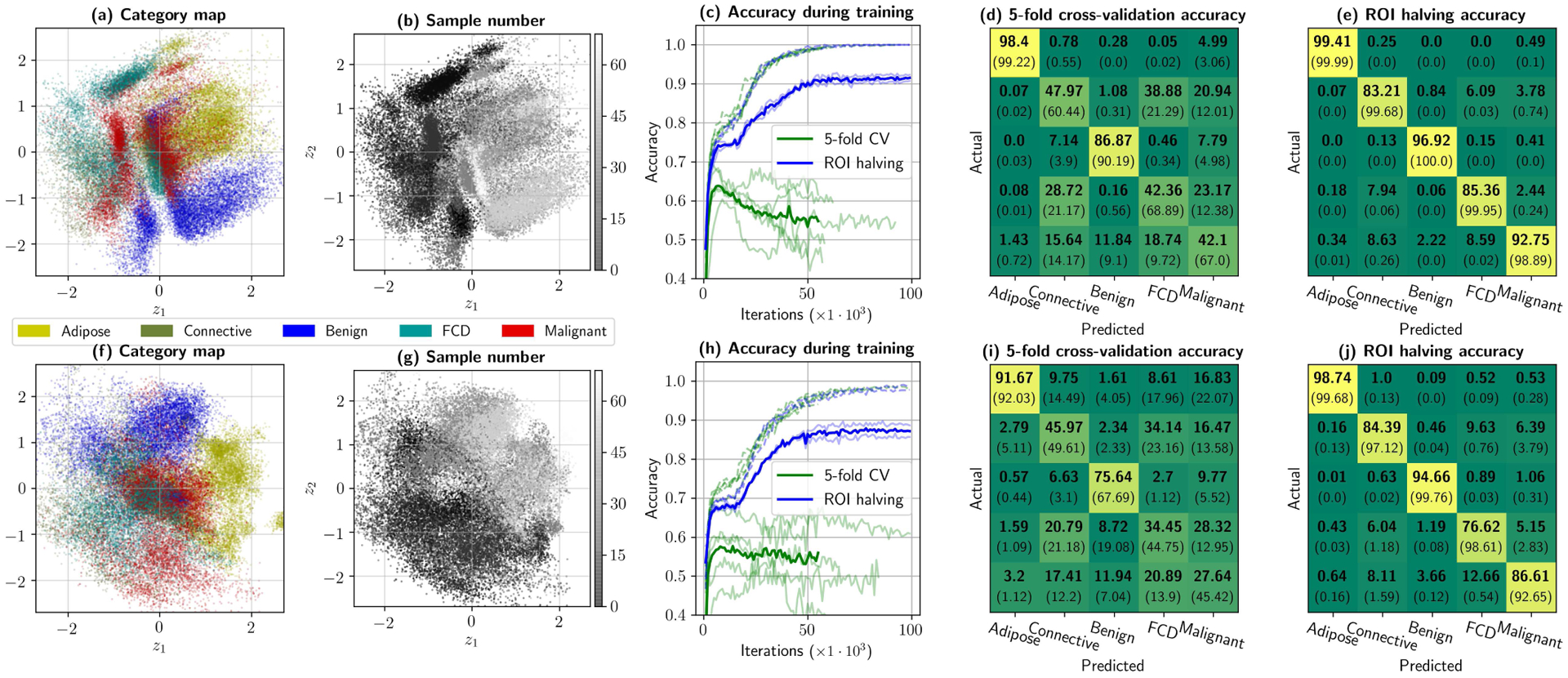
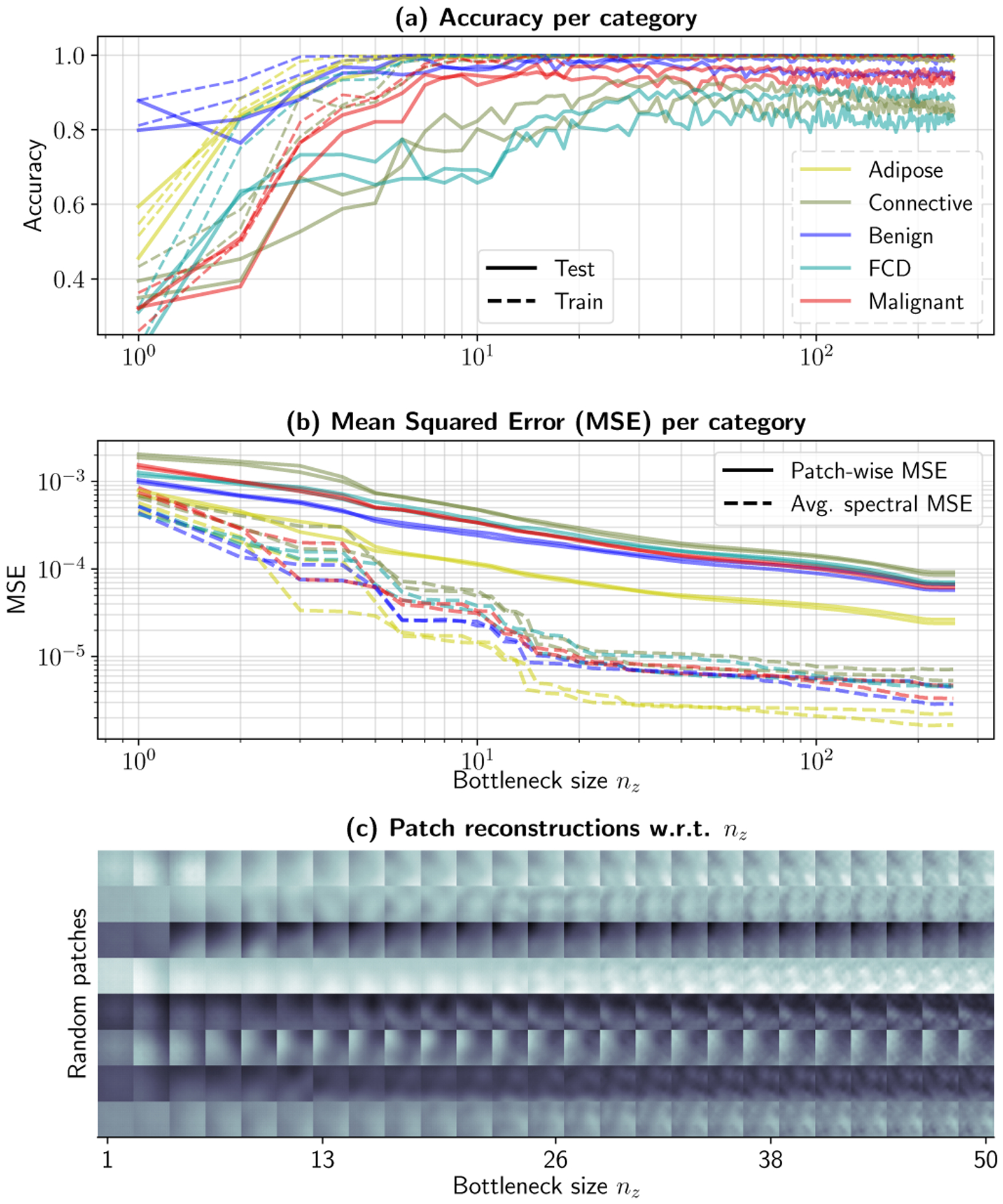
IEEE Trans Med Imaging. 2021 Jun;40(6):1687-1701. doi: 10.1109/TMI.2021.3064464. Epub 2021 Jun 1.
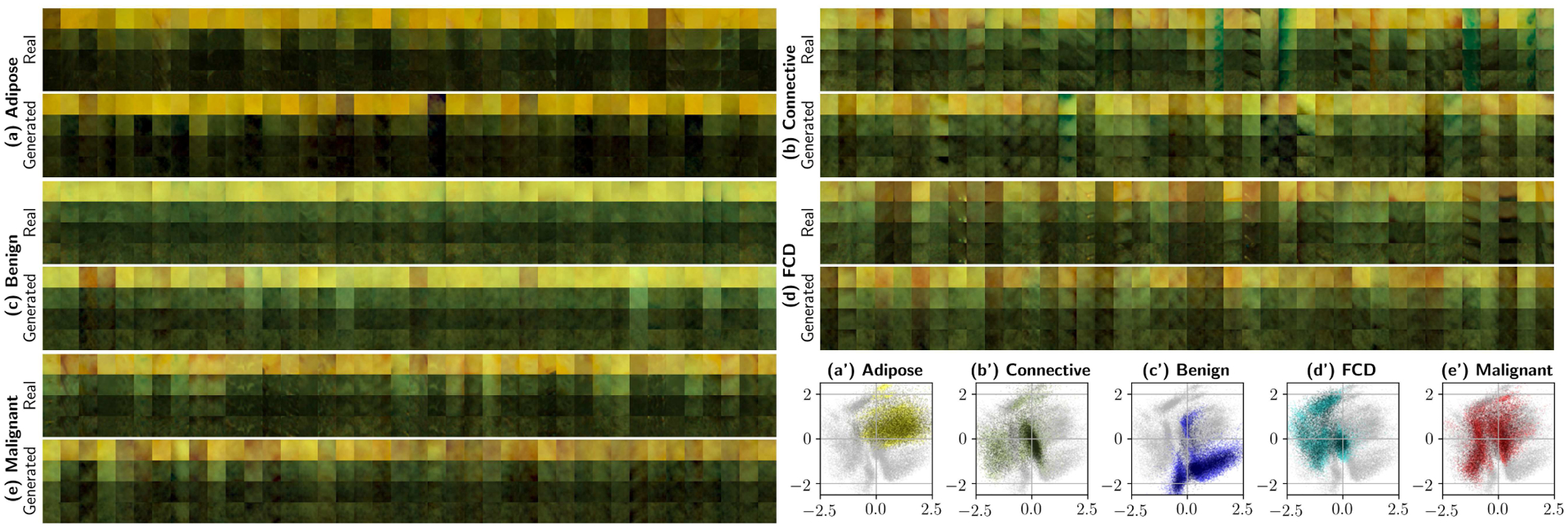
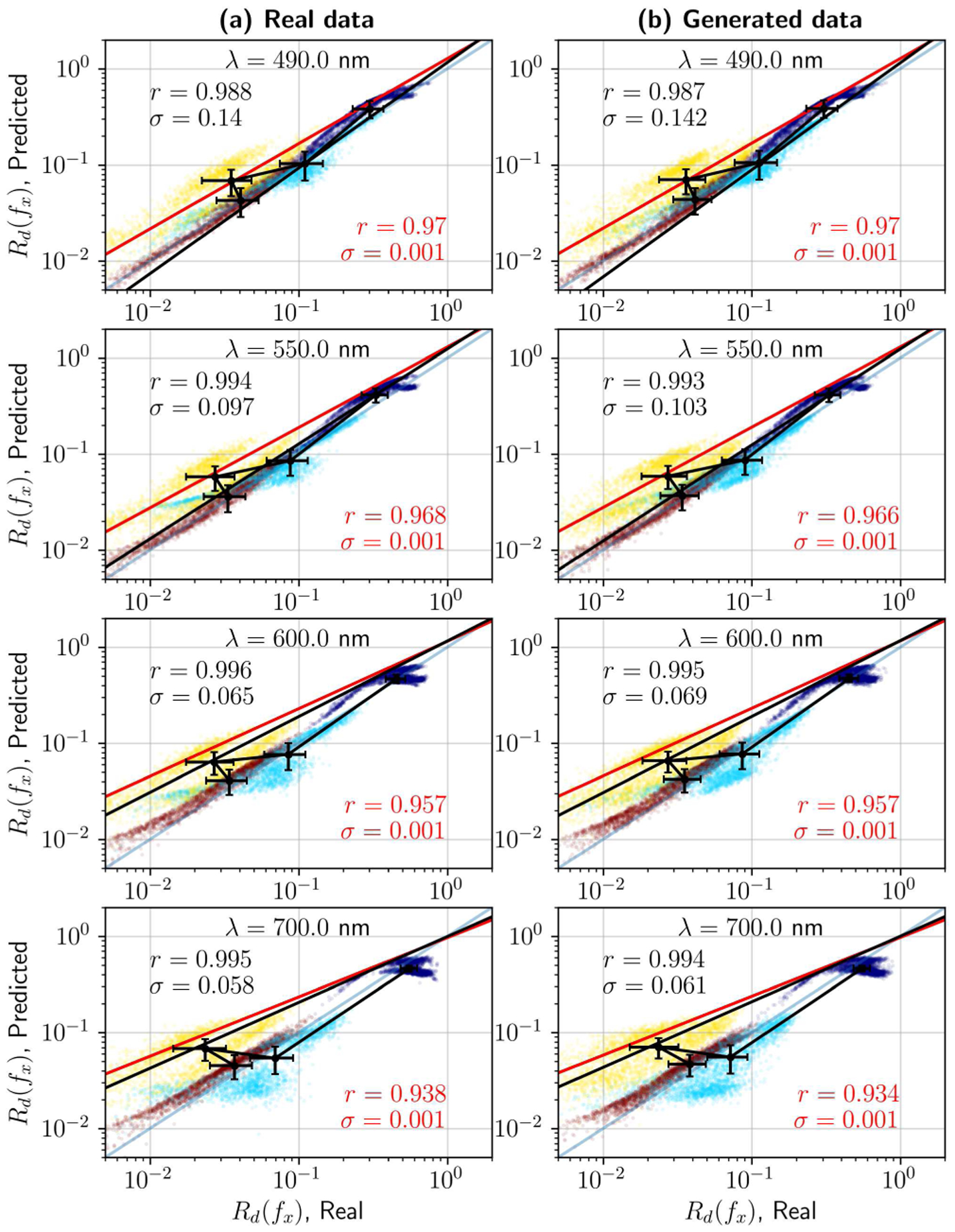
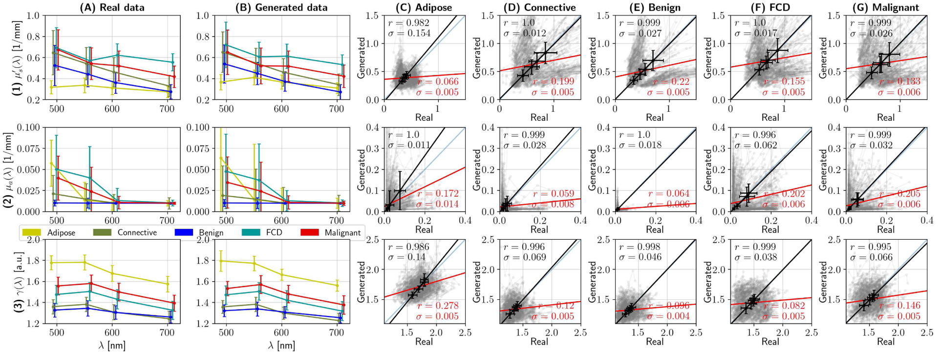
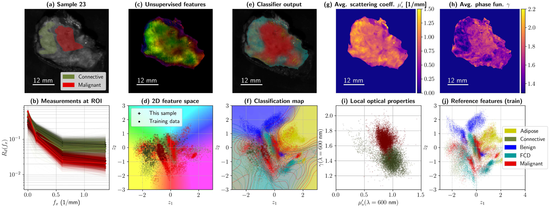
Is it possible to find deterministic relationships between optical measurements and pathophysiology in an unsupervised manner and based on data alone? Optical property quantification is a rapidly growing biomedical imaging technique for characterizing biological tissues that shows promise in a range of clinical applications, such as intraoperative breast-conserving surgery margin assessment. However, translating tissue optical properties to clinical pathology information is still a cumbersome problem due to, amongst other things, inter- and intrapatient variability, calibration, and ultimately the nonlinear behavior of light in turbid media. These challenges limit the ability of standard statistical methods to generate a simple model of pathology, requiring more advanced algorithms. We present a data-driven, nonlinear model of breast cancer pathology for real-time margin assessment of resected samples using optical properties derived from spatial frequency domain imaging data. A series of deep neural network models are employed to obtain sets of latent embeddings that relate optical data signatures to the underlying tissue pathology in a tractable manner. These self-explanatory models can translate absorption and scattering properties measured from pathology, while also being able to synthesize new data. The method was tested on a total of 70 resected breast tissue samples containing 137 regions of interest, achieving rapid optical property modeling with errors only limited by current semi-empirical models, allowing for mass sample synthesis and providing a systematic understanding of dataset properties, paving the way for deep automated margin assessment algorithms using structured light imaging or, in principle, any other optical imaging technique seeking modeling. Code is available.
是否有可能仅基于数据以无监督的方式在光学测量和病理生理学之间找到确定性关系?光学特性量化是一种快速发展的生物医学成像技术,用于对生物组织进行特征描述,在术中保乳手术边缘评估等一系列临床应用中具有广阔的前景。然而,由于个体间和个体内的变异性、校准以及光在混浊介质中的非线性行为等因素,将组织光学特性转化为临床病理信息仍然是一个繁琐的问题。这些挑战限制了标准统计方法生成简单病理模型的能力,需要更先进的算法。我们提出了一种基于数据驱动的、非线性的乳腺癌病理模型,用于使用从空间频域成像数据中获得的光学特性对切除样本进行实时边缘评估。一系列深度神经网络模型被用来获得一系列潜在的嵌入,以可处理的方式将光学数据特征与潜在的组织病理联系起来。这些自解释模型可以转换从病理学中测量的吸收和散射特性,同时也能够合成新的数据。该方法在总共 70 个切除的乳腺组织样本上进行了测试,其中包含 137 个感兴趣区域,实现了快速的光学特性建模,其误差仅受当前半经验模型的限制,允许进行大量样本合成,并提供对数据集特性的系统理解,为使用结构光成像或原则上任何其他寻求建模的光学成像技术进行深度自动边缘评估算法铺平了道路。代码可用。