• 文献检索
  • 文档翻译
  • 深度研究
  • 学术资讯
  • Suppr Zotero 插件Zotero 插件
  • 邀请有礼
  • 套餐&价格
  • 历史记录
应用&插件
Suppr Zotero 插件Zotero 插件浏览器插件Mac 客户端Windows 客户端微信小程序
定价
高级版会员购买积分包购买API积分包
服务
文献检索文档翻译深度研究API 文档MCP 服务
关于我们
关于 Suppr公司介绍联系我们用户协议隐私条款
关注我们

Suppr 超能文献

核心技术专利:CN118964589B侵权必究
粤ICP备2023148730 号-1Suppr @ 2026

文献检索

告别复杂PubMed语法,用中文像聊天一样搜索,搜遍4000万医学文献。AI智能推荐,让科研检索更轻松。

立即免费搜索

文件翻译

保留排版,准确专业,支持PDF/Word/PPT等文件格式,支持 12+语言互译。

免费翻译文档

深度研究

AI帮你快速写综述,25分钟生成高质量综述,智能提取关键信息,辅助科研写作。

立即免费体验

胎儿组织速度成像的参考值及评估胎儿心肌功能的新方法。

Reference values for fetal tissue velocity imaging and a new approach to evaluate fetal myocardial function.

作者信息

Elmstedt Nina Ne, Johnson Jonas Jj, Lind Britta Bl, Ferm-Widlund Kjerstin Kfw, Herling Lotta Lh, Westgren Magnus Mw, Brodin Lars-Åke Lab

出版信息

Cardiovasc Ultrasound. 2013 Aug 16;11:29. doi: 10.1186/1476-7120-11-29.

DOI:10.1186/1476-7120-11-29
PMID:23947791
原文链接:https://pmc.ncbi.nlm.nih.gov/articles/PMC3751897/
Abstract

OBJECTIVES

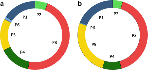
Myocardial function can be evaluated using color-coded tissue velocity imaging (TVI) to analyze the longitudinal myocardial velocity profile, and by expressing the motion of the atrioventricular plane during a cardiac cycle as coordinated events in the cardiac state diagram (CSD). The objective of this study was to establish gestational age specific reference values for fetal TVI measurements and to introduce the CSD as a potential aid in fetal myocardial evaluation.

METHODS

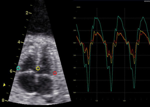
TVI recordings from 125 healthy fetuses, at 18 to 42 weeks of gestation, were performed with the transducer perpendicular to the apex to provide a four-chamber view. The myocardial velocity data was extracted from the basal segment of septum as well as the left and right ventricular free wall for subsequent offline analysis.

RESULTS

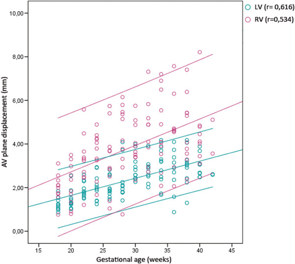
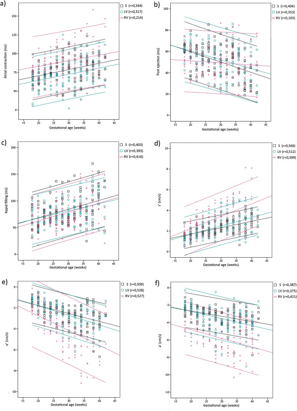
During a cardiac cycle the longitudinal peak velocities of septum increased with gestational age, as did the peak velocities of the left and right ventricular free wall, except for the peak velocity of post ejection. The duration of rapid filling and atrial contraction increased during pregnancy while the duration of post ejection decreased. The duration of pre ejection and ventricular ejection did not change significantly with gestational age.

CONCLUSION

Evaluating fetal systolic and diastolic performance using TVI together with CSD could contribute to increase the knowledge and understanding of fetal myocardial function and dysfunction. The pre and post ejection phases are the variables most likely to indicate fetuses with abnormal myocardial function.

摘要

目的

可使用彩色编码组织速度成像(TVI)评估心肌功能,以分析心肌纵向速度剖面,并通过将心动周期中房室平面的运动表示为心脏状态图(CSD)中的协调事件来进行评估。本研究的目的是建立特定孕周的胎儿TVI测量参考值,并引入CSD作为胎儿心肌评估的潜在辅助手段。

方法

对125例妊娠18至42周的健康胎儿进行TVI记录,将换能器垂直于心尖以获得四腔心视图。从室间隔基底部以及左、右心室游离壁提取心肌速度数据,以便后续离线分析。

结果

在心动周期中,除射血后期峰值速度外,室间隔纵向峰值速度以及左、右心室游离壁峰值速度均随孕周增加。妊娠期间快速充盈期和心房收缩期持续时间增加,而射血后期持续时间减少。射血前期和心室射血期持续时间随孕周无显著变化。

结论

使用TVI结合CSD评估胎儿的收缩和舒张功能,有助于增加对胎儿心肌功能和功能障碍的认识和理解。射血前期和射血后期是最有可能提示心肌功能异常胎儿的变量。

https://cdn.ncbi.nlm.nih.gov/pmc/blobs/288c/3751897/b1df6ad17d22/1476-7120-11-29-5.jpg
https://cdn.ncbi.nlm.nih.gov/pmc/blobs/288c/3751897/c12976a110cb/1476-7120-11-29-1.jpg
https://cdn.ncbi.nlm.nih.gov/pmc/blobs/288c/3751897/b391199f4af3/1476-7120-11-29-2.jpg
https://cdn.ncbi.nlm.nih.gov/pmc/blobs/288c/3751897/ab3048e2b2c1/1476-7120-11-29-3.jpg
https://cdn.ncbi.nlm.nih.gov/pmc/blobs/288c/3751897/7eda4a4afabb/1476-7120-11-29-4.jpg
https://cdn.ncbi.nlm.nih.gov/pmc/blobs/288c/3751897/b1df6ad17d22/1476-7120-11-29-5.jpg

相似文献

1
Reference values for fetal tissue velocity imaging and a new approach to evaluate fetal myocardial function.胎儿组织速度成像的参考值及评估胎儿心肌功能的新方法。
Cardiovasc Ultrasound. 2013 Aug 16;11:29. doi: 10.1186/1476-7120-11-29.
2
Automated analysis of fetal cardiac function using color tissue Doppler imaging in second half of normal pregnancy.应用彩色组织多普勒成像技术对正常妊娠后半期胎儿心功能进行自动分析。
Ultrasound Obstet Gynecol. 2019 Mar;53(3):348-357. doi: 10.1002/uog.19037.
3
Automated quantitative evaluation of fetal atrioventricular annular plane systolic excursion.胎儿房室环平面收缩期位移的自动定量评估
Ultrasound Obstet Gynecol. 2021 Dec;58(6):853-863. doi: 10.1002/uog.23703.
4
Evaluation of ventricular myocardial velocities and heart motion of the fetal heart by tissue Doppler image.应用组织多普勒成像技术评估胎儿心脏的心室心肌速度和心脏运动。
J Med Assoc Thai. 2001 Aug;84(8):1158-63.
5
Fetal cardiac muscle contractility decreases with gestational age: a color-coded tissue velocity imaging study.胎儿心肌收缩力随孕周增加而降低:一项彩色编码组织速度成像研究。
Cardiovasc Ultrasound. 2012 May 9;10:19. doi: 10.1186/1476-7120-10-19.
6
Quantification of regional left and right ventricular longitudinal function in 75 normal fetuses using ultrasound-based strain rate and strain imaging.使用基于超声的应变率和应变成像技术对75例正常胎儿的左右心室局部纵向功能进行量化分析。
Ultrasound Med Biol. 2005 Sep;31(9):1159-62. doi: 10.1016/j.ultrasmedbio.2005.05.011.
7
Temporal frequency requirements for tissue velocity imaging of the fetal heart.胎儿心脏组织速度成像的时间频率要求。
Ultrasound Obstet Gynecol. 2011 Oct;38(4):413-7. doi: 10.1002/uog.8872.
8
Tissue Doppler imaging of the fetal heart.胎儿心脏的组织多普勒成像。
Ultrasound Obstet Gynecol. 2000 Nov;16(6):530-5. doi: 10.1046/j.1469-0705.2000.00251.x.
9
Regional atrial myocardial velocity in normal fetuses: evaluation by quantitative tissue velocity imaging.
Echocardiography. 2012 Feb;29(2):182-6. doi: 10.1111/j.1540-8175.2011.01562.x. Epub 2011 Nov 8.
10
Analysis of segmental and global function of the fetal heart using novel automatic functional imaging.使用新型自动功能成像技术分析胎儿心脏的节段性和整体功能。
J Am Soc Echocardiogr. 2008 Feb;21(2):146-50. doi: 10.1016/j.echo.2007.05.007. Epub 2007 Jul 12.

引用本文的文献

1
Fetal cardiac function at intrauterine transfusion assessed by automated analysis of color tissue Doppler recordings.通过自动分析彩色组织多普勒记录评估宫内输血胎儿的心脏功能。
Cardiovasc Ultrasound. 2020 Aug 13;18(1):34. doi: 10.1186/s12947-020-00214-1.
2
Impact of maternal diabetes and obesity on fetal cardiac functions.母亲糖尿病和肥胖对胎儿心脏功能的影响。
Egypt Heart J. 2020 Jul 31;72(1):46. doi: 10.1186/s43044-020-00077-x.
3
Measurement of cardiac function by cardiac time intervals, applicability in normal pregnancy and twin-to-twin transfusion syndrome.

本文引用的文献

1
The cardiac state diagram as a novel approach for the evaluation of pre- and post-ejection phases of the cardiac cycle in asphyxiated fetal lambs.心脏状态图作为一种评估窒息胎儿羊心动周期射血前期和射血后期的新方法。
Ultrasound Med Biol. 2013 Sep;39(9):1682-7. doi: 10.1016/j.ultrasmedbio.2013.04.012. Epub 2013 Jul 9.
2
Second trimester ultrasound: reference values for two-dimensional speckle tracking-derived longitudinal strain, strain rate and time to peak deformation of the fetal heart.中孕期超声:二维斑点追踪技术衍生的胎儿心脏纵向应变、应变率和达峰时间的参考值。
J Am Soc Echocardiogr. 2012 Dec;25(12):1333-41. doi: 10.1016/j.echo.2012.09.011.
3
通过心脏时间间期测量心功能,在正常妊娠及双胎输血综合征中的适用性。
J Echocardiogr. 2019 Sep;17(3):129-137. doi: 10.1007/s12574-018-0401-9. Epub 2018 Oct 20.
4
Maternal vitamin D deficiency and fetal distress/birth asphyxia: a population-based nested case-control study.母亲维生素D缺乏与胎儿窘迫/出生窒息:一项基于人群的巢式病例对照研究。
BMJ Open. 2016 Sep 22;6(9):e009733. doi: 10.1136/bmjopen-2015-009733.
5
Contractile properties of developing human fetal cardiac muscle.发育中的人类胎儿心肌的收缩特性。
J Physiol. 2016 Jan 15;594(2):437-52. doi: 10.1113/JP271290. Epub 2015 Dec 7.
Feasibility and reproducibility of a standard protocol for 2D speckle tracking and tissue Doppler-based strain and strain rate analysis of the fetal heart.
二维斑点追踪和组织多普勒应变及应变率分析胎儿心脏的标准方案的可行性和可重复性。
Fetal Diagn Ther. 2012;32(1-2):96-108. doi: 10.1159/000337329. Epub 2012 Jun 19.
4
Evaluation of conventional Doppler fetal cardiac function parameters: E/A ratios, outflow tracts, and myocardial performance index.评估常规多普勒胎儿心功能参数:E/A 比值、流出道和心肌做功指数。
Fetal Diagn Ther. 2012;32(1-2):22-9. doi: 10.1159/000330792. Epub 2012 Jun 1.
5
Fetal color-coded tissue Doppler echocardiography: experiences with the "tracking method" in normal fetuses.胎儿彩色组织多普勒超声心动图:“跟踪法”在正常胎儿中的应用经验。
Eur J Obstet Gynecol Reprod Biol. 2012 Sep;164(1):10-4. doi: 10.1016/j.ejogrb.2012.05.013. Epub 2012 May 30.
6
Temporal frequency requirements for tissue velocity imaging of the fetal heart.胎儿心脏组织速度成像的时间频率要求。
Ultrasound Obstet Gynecol. 2011 Oct;38(4):413-7. doi: 10.1002/uog.8872.
7
The importance of venous Doppler velocimetry for evaluation of intrauterine growth restriction.静脉多普勒速度测定在评估宫内生长受限中的重要性。
J Ultrasound Med. 2011 Apr;30(4):529-45. doi: 10.7863/jum.2011.30.4.529.
8
Is fetal cardiac function gender dependent?胎儿心脏功能是否存在性别差异?
Prenat Diagn. 2011 Jun;31(6):536-42. doi: 10.1002/pd.2738. Epub 2011 Mar 17.
9
Color Doppler myocardial imaging demonstrates reduced diastolic tissue velocity in growth retarded fetuses with flow redistribution.彩色多普勒心肌成像显示血流重新分布的生长受限胎儿舒张期组织速度降低。
Eur J Obstet Gynecol Reprod Biol. 2011 Apr;155(2):140-5. doi: 10.1016/j.ejogrb.2010.12.020. Epub 2011 Jan 21.
10
Temporal and spatial performance of vector velocity imaging in the human fetal heart.胎儿心脏向量速度成像的时-空间性能。
Ultrasound Obstet Gynecol. 2011 Feb;37(2):150-7. doi: 10.1002/uog.8815.