Department of Physical Therapy and Human Movement Sciences, Feinberg School of Medicine, Northwestern University, Chicago, IL, USA.
Osteoarthritis Cartilage. 2013 Nov;21(11):1668-73. doi: 10.1016/j.joca.2013.08.007. Epub 2013 Aug 12.
Varus thrust visualized during walking is associated with a greater medial knee load and an increased risk of medial knee osteoarthritis (OA) progression. Little is known about how varus thrust presence determined by visual observation relates to quantitative gait kinematic data. We hypothesized that varus thrust presence is associated with greater knee frontal plane dynamic movement during the stance phase of gait.
Participants had knee OA in at least one knee. Trained examiners assessed participants for varus thrust presence during ambulation. Frontal plane knee motion during ambulation was captured using external passive reflective markers and an 8-camera motion analysis system. To examine the cross-sectional relationship between varus thrust and frontal plane knee motion, we used multivariable regression models with the quantitative motion measures as dependent variables and varus thrust (present/absent) as predictor; models were adjusted for age, gender, body mass index (BMI), gait speed, and knee static alignment.
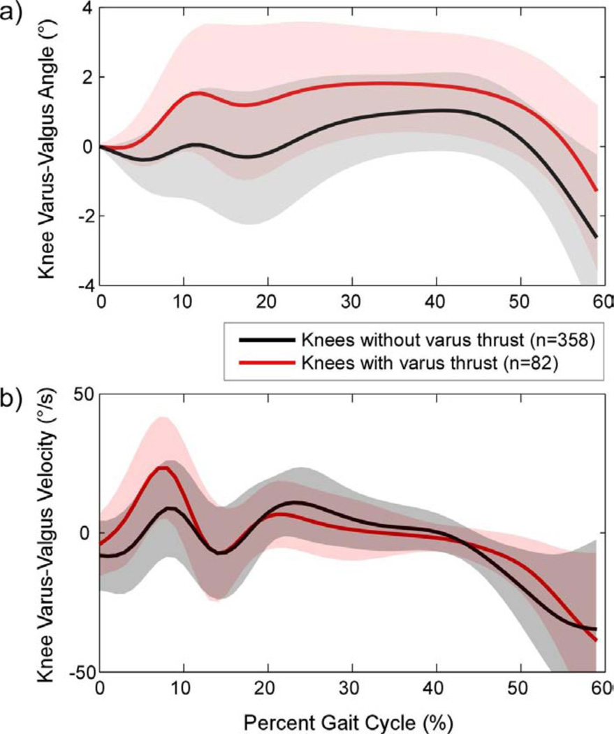
236 persons [mean BMI: 28.5 kg/m(2) (standard deviation (SD) 5.5), mean age: 64.9 years (SD 10.4), 75.8% women] contributing 440 knees comprised the study sample. 82 knees (18.6%) had definite varus thrust. Knees with varus thrust had greater peak varus angle and greater peak varus angular velocity during stance than knees without varus thrust (mean differences 0.90° and 6.65°/s, respectively). These patterns remained significant after adjusting for age, gender, BMI, gait speed, and knee static alignment.
Visualized varus thrust during walking was associated with a greater peak knee varus angular velocity and a greater peak knee varus angle during stance phase of gait.
行走时出现的内翻推力与更大的内侧膝关节负荷和内侧膝关节骨关节炎(OA)进展的风险增加有关。对于通过视觉观察确定的内翻推力的存在与定量步态运动学数据之间的关系知之甚少。我们假设,在步态的站立阶段,内翻推力的存在与更大的膝关节额状面动态运动有关。
参与者至少有一侧膝关节 OA。受过训练的检查者在行走时评估参与者是否存在内翻推力。使用外部无源反射标记和 8 个摄像机运动分析系统来捕获行走时的额状面膝关节运动。为了研究内翻推力与额状面膝关节运动之间的横断面关系,我们使用多变量回归模型,将定量运动测量值作为因变量,内翻推力(存在/不存在)作为预测因子;模型调整了年龄、性别、体重指数(BMI)、步态速度和膝关节静态排列。
236 名参与者(平均 BMI:28.5 kg/m²(标准差(SD)5.5),平均年龄:64.9 岁(SD 10.4),75.8%为女性)贡献了 440 个膝关节,构成了研究样本。82 个膝关节(18.6%)存在明确的内翻推力。与没有内翻推力的膝关节相比,具有内翻推力的膝关节在站立时具有更大的峰值内翻角度和更大的峰值内翻角速度(平均差异分别为 0.90°和 6.65°/s)。这些模式在调整年龄、性别、BMI、步态速度和膝关节静态排列后仍然显著。
行走时出现的可视化内翻推力与站立阶段膝关节更大的峰值内翻角速度和更大的峰值内翻角度有关。