Cimino Patrick J, Perrin Richard J
Department of Pathology and Immunology, Division of Neuropathology, Washington University School of Medicine, St Louis, MO.
Appl Immunohistochem Mol Morphol. 2014 Jul;22(6):442-8. doi: 10.1097/PAI.0b013e318294ca46.
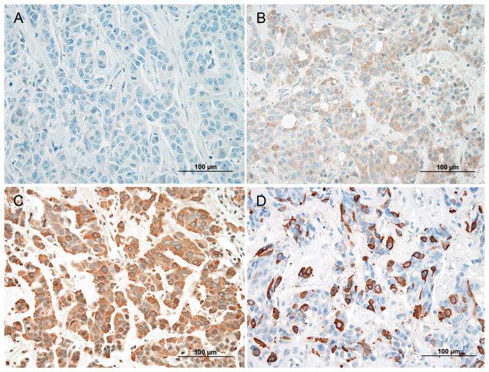
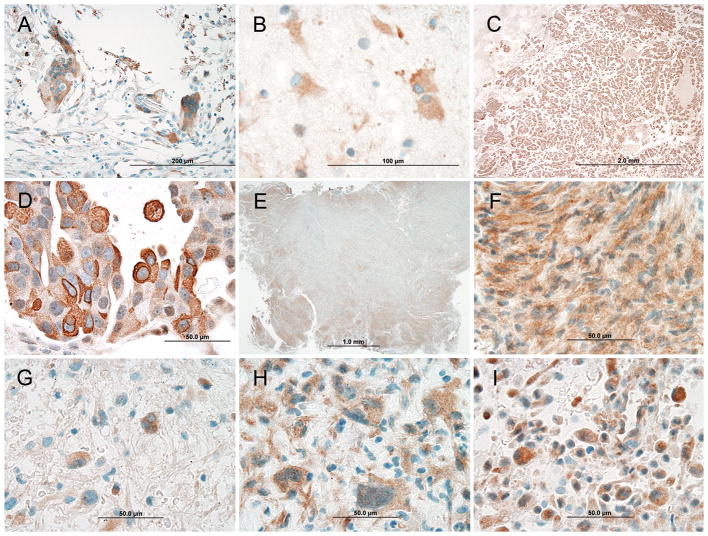
Metastases represent the most common type of intracranial neoplasm. In women, 30% of such tumors derive from breast carcinoma. In neurosurgical cases with ambiguous cellular morphology and/or limited biopsy material, immunohistochemistry (IHC) is often performed to distinguish metastases from primary central nervous system (CNS) neoplasms. IHC for mammaglobin-A (MGA), a protein expressed in a majority of breast carcinomas, is commonly applied in this setting, but its utility for distinguishing primary CNS neoplasms from metastatic breast carcinoma is unknown; the reactivity of MGA in primary and metastatic CNS neoplasms has never been described. Here, we describe the frequency and patterns of IHC reactivity for MGA in metastatic and primary CNS neoplasms from patients with well-documented histories of breast carcinoma. Following a published protocol previously applied to non-CNS neoplasms, MGA staining of moderate to strong intensity within 5% or more of a neoplasm was considered positive. On the basis of these criteria, 3 of 12 (25.0%) glioblastomas, 1 of 10 (10.0%) meningiomas, and 47 of 95 (49.5%) metastases were positive. Importantly, the cytoarchitectural staining characteristics among all 4 MGA-positive primary CNS neoplasms (cytoplasmic and nuclear) differed from those of the metastases (cytoplasmic and membranous). These findings suggest that MGA IHC staining intensity and distribution can distinguish metastases from primary CNS neoplasms (P=0.0086) in women with a history of breast carcinoma but also indicate that cytologic staining patterns must be interpreted for more accurate tumor classification.
转移瘤是颅内肿瘤最常见的类型。在女性中,此类肿瘤的30%源自乳腺癌。在细胞形态不明确和/或活检材料有限的神经外科病例中,常进行免疫组织化学(IHC)检查以区分转移瘤与原发性中枢神经系统(CNS)肿瘤。乳腺珠蛋白-A(MGA)是一种在大多数乳腺癌中表达的蛋白质,在这种情况下常用其进行免疫组化检测,但其在区分原发性CNS肿瘤与转移性乳腺癌方面的效用尚不清楚;MGA在原发性和转移性CNS肿瘤中的反应性从未被描述过。在此,我们描述了有充分记录的乳腺癌病史患者的转移性和原发性CNS肿瘤中MGA免疫组化反应性的频率和模式。按照先前应用于非CNS肿瘤的已发表方案,肿瘤内5%或更多区域出现中度至强强度的MGA染色被视为阳性。基于这些标准,12例胶质母细胞瘤中有3例(25.0%)、10例脑膜瘤中有1例(10.0%)以及95例转移瘤中有47例(49.5%)呈阳性。重要的是,所有4例MGA阳性原发性CNS肿瘤的细胞结构染色特征(细胞质和细胞核)与转移瘤(细胞质和细胞膜)不同。这些发现表明,MGA免疫组化染色强度和分布可在有乳腺癌病史的女性中区分转移瘤与原发性CNS肿瘤(P = 0.0086),但也表明必须解读细胞学染色模式以进行更准确的肿瘤分类。