Department of Chemical Engineering and ‡Department of Biomedical Engineering, University of Rochester , Rochester, New York 14627, United States.
Langmuir. 2013 Oct 1;29(39):12183-93. doi: 10.1021/la400971g. Epub 2013 Sep 17.
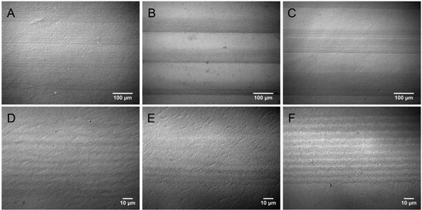
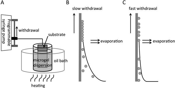
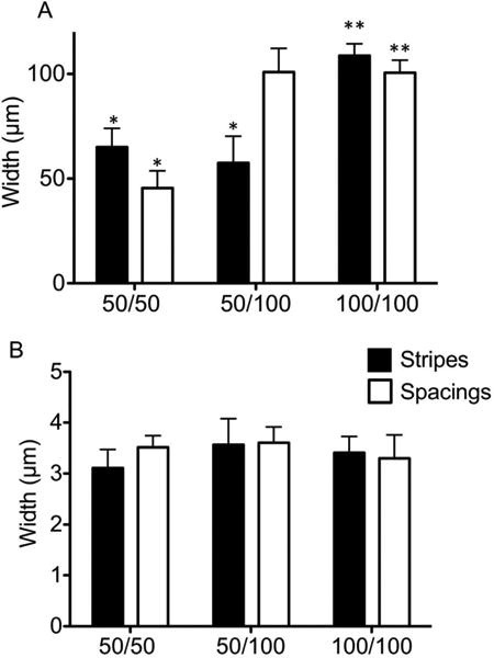
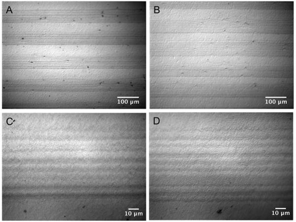
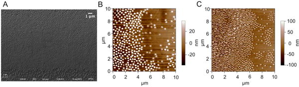
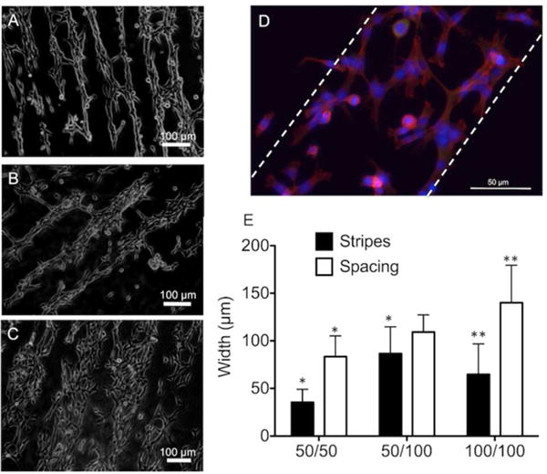
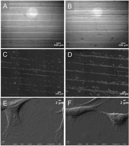
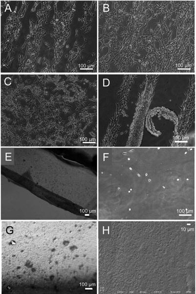
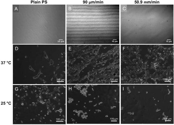
Thermoresponsive poly(N-isopropyl acrylamide) (PNIPAM) microgels were patterned on polystyrene substrates via dip coating, creating cytocompatible substrates that provided spatial control over cell adhesion. This simple dip-coating method, which exploits variable substrate withdrawal speeds forming particle suspension stripes of densely packed PNIPAM microgels, while spacings between the stripes contained sparsely distributed PNIPAM microgels. The assembly of three different PNIPAM microgel patterns, namely, patterns composed of 50 μm stripe/50 μm spacing, 50 μm stripe/100 μm spacing, and 100 μm stripe/100 μm spacing, was verified using high-resolution optical micrographs and ImageJ analysis. PNIPAM microgels existed as monolayers within stripes and spacings, as revealed by atomic force microscopy (AFM). Upon cell seeding on PNIPAM micropatterned substrates, NIH3T3 fibroblast cells preferentially adhered within spacings to form cell patterns. Three days after cell seeding, cells proliferated to form confluent cell layers. The thermoresponsiveness of the underlying PNIPAM microgels was then utilized to recover fibroblast cell sheets from substrates simply by lowering the temperature without disrupting the underlying PNIPAM microgel patterns. Harvested cell sheets similar to these have been used for multiple tissue engineering applications. Also, this simple, low-cost, template-free dip-coating technique can be utilized to micropattern multifunctional PNIPAM microgels, generating complex stimuli-responsive substrates to study cell-material interactions and allow drug delivery to cells in a spatially and temporally controlled manner.
温敏性聚(N-异丙基丙烯酰胺)(PNIPAM)微凝胶通过浸涂在聚苯乙烯基底上形成图案,构建了细胞相容的基底,实现了对细胞黏附的空间控制。这种简单的浸涂方法利用了可变的基底撤出速度,形成了紧密堆积的 PNIPAM 微凝胶的颗粒悬浮条纹,而条纹之间的间隔则包含稀疏分布的 PNIPAM 微凝胶。通过高分辨率光学显微镜和 ImageJ 分析,验证了三种不同的 PNIPAM 微凝胶图案的组装,即由 50μm 条纹/50μm 间隔、50μm 条纹/100μm 间隔和 100μm 条纹/100μm 间隔组成的图案。原子力显微镜(AFM)揭示了 PNIPAM 微凝胶存在于条纹和间隔内的单层结构。在将 NIH3T3 成纤维细胞接种到 PNIPAM 微图案化基底上后,细胞优先在间隔内黏附形成细胞图案。细胞接种 3 天后,细胞增殖形成细胞层。然后,通过降低温度而不破坏下面的 PNIPAM 微凝胶图案,利用下面的 PNIPAM 微凝胶的温敏性,简单地从基底上回收成纤维细胞片。从基底上回收的细胞片类似于这些细胞片已经用于多种组织工程应用。此外,这种简单、低成本、无模板的浸涂技术可以用于微图案化多功能 PNIPAM 微凝胶,生成复杂的刺激响应性基底,以研究细胞-材料相互作用,并允许以时空控制的方式向细胞输送药物。