Department of Biosciences and Informatics, Faculty of Science and Technology, Keio University, Kohoku-ku, Yokohama, Kanagawa, Japan.
PLoS One. 2013 Aug 20;8(8):e71739. doi: 10.1371/journal.pone.0071739. eCollection 2013.
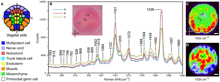
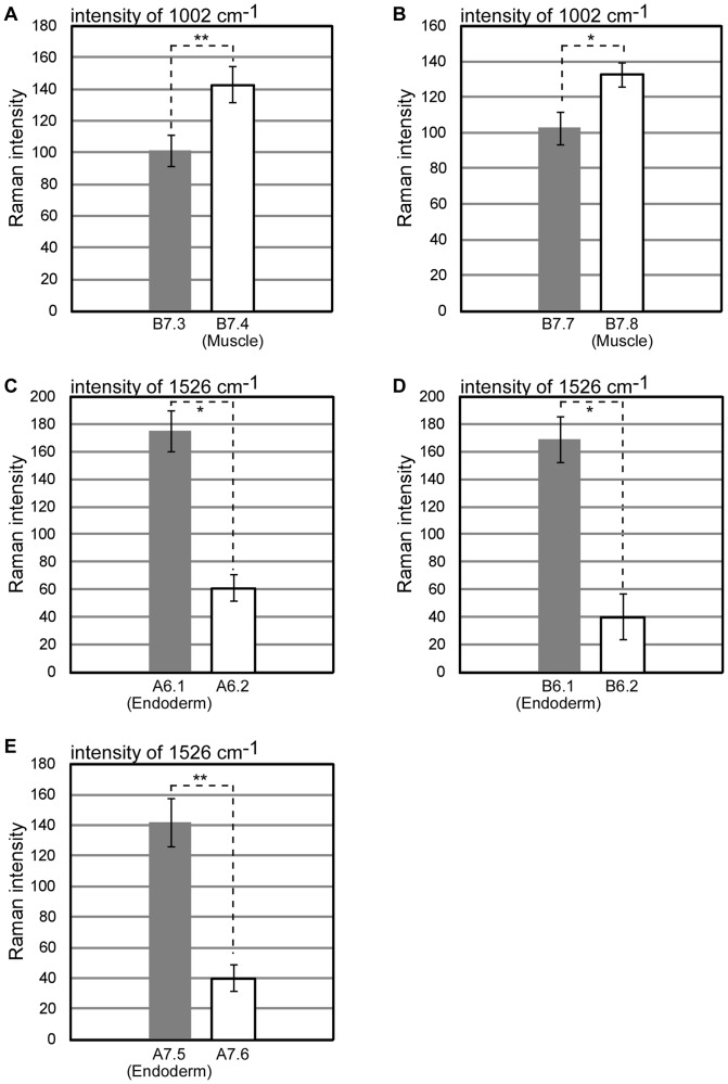
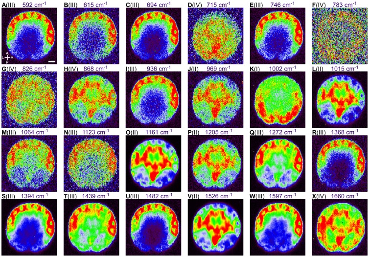
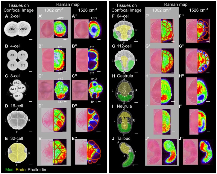
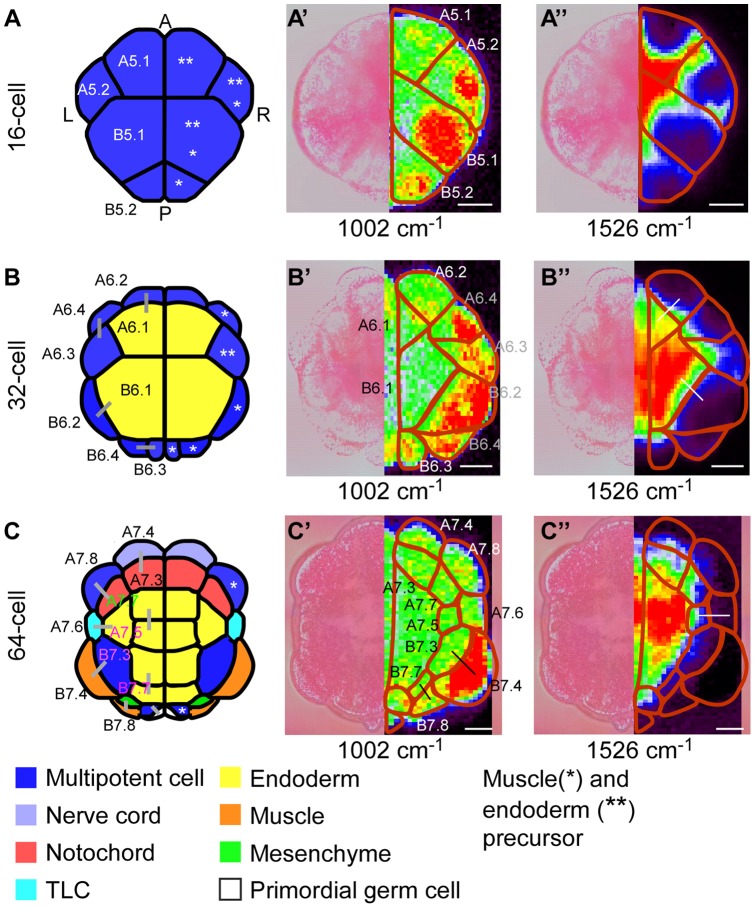
Intracellular composition and the distribution of bio-molecules play central roles in the specification of cell fates and morphogenesis during embryogenesis. Consequently, investigation of changes in the expression and distribution of bio-molecules, especially mRNAs and proteins, is an important challenge in developmental biology. Raman spectroscopic imaging, a non-invasive and label-free technique, allows simultaneous imaging of the intracellular composition and distribution of multiple bio-molecules. In this study, we explored the application of Raman spectroscopic imaging in the whole Ciona intestinalis embryo during development. Analysis of Raman spectra scattered from C. intestinalis embryos revealed a number of localized patterns of high Raman intensity within the embryo. Based on the observed distribution of bio-molecules, we succeeded in identifying the location and structure of differentiated muscle and endoderm within the whole embryo, up to the tailbud stage, in a label-free manner. Furthermore, during cell differentiation, we detected significant differences in cell state between muscle/endoderm daughter cells and daughter cells with other fates that had divided from the same mother cells; this was achieved by focusing on the Raman intensity of single Raman bands at 1002 or 1526 cm(-1), respectively. This study reports the first application of Raman spectroscopic imaging to the study of identifying and characterizing differentiating tissues in a whole chordate embryo. Our results suggest that Raman spectroscopic imaging is a feasible label-free technique for investigating the developmental process of the whole embryo of C. intestinalis.
细胞内组成和生物分子的分布在胚胎发生过程中细胞命运的特化和形态发生中起着核心作用。因此,研究生物分子(特别是 mRNA 和蛋白质)的表达和分布的变化是发育生物学中的一个重要挑战。拉曼光谱成像技术是一种非侵入性和无标记的技术,可同时对细胞内多种生物分子的组成和分布进行成像。在本研究中,我们探讨了拉曼光谱成像技术在整个海鞘胚胎发育过程中的应用。对海鞘胚胎散射的拉曼光谱进行分析,揭示了胚胎内存在许多局域化的高强度拉曼区域。基于观察到的生物分子分布,我们成功地以无标记的方式识别了整个胚胎中分化的肌肉和内胚层的位置和结构,直到尾芽阶段。此外,在细胞分化过程中,我们检测到肌肉/内胚层子细胞和来自同一母细胞的具有其他命运的子细胞之间的细胞状态存在显著差异;这是通过分别聚焦于 1002 或 1526 cm(-1)处的单个拉曼带的拉曼强度来实现的。本研究首次报道了将拉曼光谱成像技术应用于鉴定和表征整个脊索动物胚胎中分化组织的研究。我们的结果表明,拉曼光谱成像技术是一种可行的无标记技术,可用于研究海鞘胚胎的整个发育过程。