• 文献检索
  • 文档翻译
  • 深度研究
  • 学术资讯
  • Suppr Zotero 插件Zotero 插件
  • 邀请有礼
  • 套餐&价格
  • 历史记录
应用&插件
Suppr Zotero 插件Zotero 插件浏览器插件Mac 客户端Windows 客户端微信小程序
定价
高级版会员购买积分包购买API积分包
服务
文献检索文档翻译深度研究API 文档MCP 服务
关于我们
关于 Suppr公司介绍联系我们用户协议隐私条款
关注我们

Suppr 超能文献

核心技术专利:CN118964589B侵权必究
粤ICP备2023148730 号-1Suppr @ 2026

文献检索

告别复杂PubMed语法,用中文像聊天一样搜索,搜遍4000万医学文献。AI智能推荐,让科研检索更轻松。

立即免费搜索

文件翻译

保留排版,准确专业,支持PDF/Word/PPT等文件格式,支持 12+语言互译。

免费翻译文档

深度研究

AI帮你快速写综述,25分钟生成高质量综述,智能提取关键信息,辅助科研写作。

立即免费体验

基于金纳米粒子的表面增强拉曼散射用于胚胎干细胞分化的无创分子探测。

Gold nanoparticle-based surface-enhanced Raman scattering for noninvasive molecular probing of embryonic stem cell differentiation.

机构信息

Department of Applied Physics, Graduate School of Engineering, Osaka University, Suita City, Osaka, Japan.

出版信息

PLoS One. 2011;6(8):e22802. doi: 10.1371/journal.pone.0022802. Epub 2011 Aug 4.

DOI:10.1371/journal.pone.0022802
PMID:21829653
原文链接:https://pmc.ncbi.nlm.nih.gov/articles/PMC3150363/
Abstract

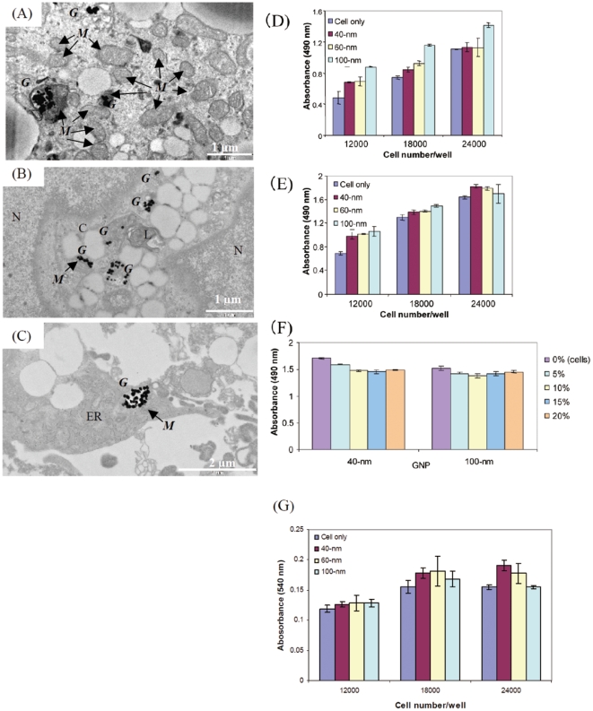
This study reports the use of gold nanoparticle-based surface-enhanced Raman scattering (SERS) for probing the differentiation of mouse embryonic stem (mES) cells, including undifferentiated single cells, embryoid bodies (EBs), and terminally differentiated cardiomyocytes. Gold nanoparticles (GNPs) were successfully delivered into all 3 mES cell differentiation stages without affecting cell viability or proliferation. Transmission electron microscopy (TEM) confirmed the localization of GNPs inside the following cell organelles: mitochondria, secondary lysosome, and endoplasmic reticulum. Using bright- and dark-field imaging, the bright scattering of GNPs and nanoaggregates in all 3 ES cell differentiation stages could be visualized. EB (an early differentiation stage) and terminally differentiated cardiomyocytes both showed SERS peaks specific to metabolic activity in the mitochondria and to protein translation (amide I, amide II, and amide III peaks). These peaks have been rarely identified in undifferentiated single ES cells. Spatiotemporal changes observed in the SERS spectra from terminally differentiated cardiomyocyte tissues revealed local and dynamic molecular interactions as well as transformations during ES cell differentiation.

摘要

本研究报告了基于金纳米粒子的表面增强拉曼散射(SERS)在探测小鼠胚胎干细胞(mES 细胞)分化中的应用,包括未分化的单细胞、类胚体(EB)和终末分化的心肌细胞。金纳米粒子(GNPs)成功地递送至所有 3 个 mES 细胞分化阶段,而不影响细胞活力或增殖。透射电子显微镜(TEM)证实了 GNPs 定位于以下细胞细胞器内:线粒体、次级溶酶体和内质网。使用明场和暗场成像,可以观察到所有 3 个 ES 细胞分化阶段中 GNPs 和纳米聚集体的明亮散射。EB(早期分化阶段)和终末分化的心肌细胞都显示出与线粒体代谢活性和蛋白质翻译(酰胺 I、酰胺 II 和酰胺 III 峰)相关的特异性 SERS 峰。这些峰在未分化的单细胞 ES 细胞中很少被识别。终末分化的心肌细胞组织的 SERS 光谱中观察到的时空变化揭示了 ES 细胞分化过程中局部和动态的分子相互作用以及转化。

https://cdn.ncbi.nlm.nih.gov/pmc/blobs/967c/3150363/6610f976a9a4/pone.0022802.g006.jpg
https://cdn.ncbi.nlm.nih.gov/pmc/blobs/967c/3150363/e08103597cfb/pone.0022802.g001.jpg
https://cdn.ncbi.nlm.nih.gov/pmc/blobs/967c/3150363/4528f17cdff9/pone.0022802.g002.jpg
https://cdn.ncbi.nlm.nih.gov/pmc/blobs/967c/3150363/668971332825/pone.0022802.g003.jpg
https://cdn.ncbi.nlm.nih.gov/pmc/blobs/967c/3150363/52a29c7db8a5/pone.0022802.g004.jpg
https://cdn.ncbi.nlm.nih.gov/pmc/blobs/967c/3150363/c37a22e022bc/pone.0022802.g005.jpg
https://cdn.ncbi.nlm.nih.gov/pmc/blobs/967c/3150363/6610f976a9a4/pone.0022802.g006.jpg

相似文献

1
Gold nanoparticle-based surface-enhanced Raman scattering for noninvasive molecular probing of embryonic stem cell differentiation.基于金纳米粒子的表面增强拉曼散射用于胚胎干细胞分化的无创分子探测。
PLoS One. 2011;6(8):e22802. doi: 10.1371/journal.pone.0022802. Epub 2011 Aug 4.
2
Gold nanocage-based surface-enhanced Raman scattering probes for long-term monitoring of intracellular microRNA during bone marrow stem cell differentiation.基于金纳米笼的表面增强拉曼散射探针用于长期监测骨髓干细胞分化过程中的细胞内 microRNA。
Nanoscale. 2020 Jan 23;12(3):1513-1527. doi: 10.1039/c9nr07791j.
3
3D graphene oxide-encapsulated gold nanoparticles to detect neural stem cell differentiation.3D 石墨烯氧化物包裹的金纳米粒子用于检测神经干细胞分化。
Biomaterials. 2013 Nov;34(34):8660-70. doi: 10.1016/j.biomaterials.2013.07.101. Epub 2013 Aug 12.
4
Gold Nanoparticles in Single-Cell Analysis for Surface Enhanced Raman Scattering.用于表面增强拉曼散射的单细胞分析中的金纳米颗粒
Molecules. 2016 Nov 25;21(12):1617. doi: 10.3390/molecules21121617.
5
Relating surface-enhanced Raman scattering signals of cells to gold nanoparticle aggregation as determined by LA-ICP-MS micromapping.通过激光烧蚀电感耦合等离子体质谱微成像确定细胞的表面增强拉曼散射信号与金纳米颗粒聚集的关系。
Anal Bioanal Chem. 2014 Nov;406(27):7003-14. doi: 10.1007/s00216-014-8069-0. Epub 2014 Aug 14.
6
3D SERS (surface enhanced Raman scattering) imaging of intracellular pathways.细胞内信号通路的三维表面增强拉曼散射成像
Methods. 2014 Jul 1;68(2):348-53. doi: 10.1016/j.ymeth.2014.02.007. Epub 2014 Feb 17.
7
Surface-enhanced Raman scattering in local optical fields of silver and gold nanoaggregates-from single-molecule Raman spectroscopy to ultrasensitive probing in live cells.银和金纳米聚集体局部光学场中的表面增强拉曼散射——从单分子拉曼光谱到活细胞中的超灵敏探测
Acc Chem Res. 2006 Jul;39(7):443-50. doi: 10.1021/ar050107x.
8
Intracellular surface-enhanced Raman scattering probes based on TAT peptide-conjugated Au nanostars for distinguishing the differentiation of lung resident mesenchymal stem cells.基于 TAT 肽修饰的金纳米星的细胞内表面增强拉曼散射探针,用于区分肺驻留间充质干细胞的分化。
Biomaterials. 2015 Jul;58:10-25. doi: 10.1016/j.biomaterials.2015.04.010. Epub 2015 Apr 29.
9
Bio-hybrid gold nanoparticles as SERS probe for rapid bacteria cell identification.生物杂化金纳米粒子作为 SERS 探针用于快速细菌细胞鉴定。
Spectrochim Acta A Mol Biomol Spectrosc. 2020 Jan 5;224:117394. doi: 10.1016/j.saa.2019.117394. Epub 2019 Jul 16.
10
Raman imaging diagnosis of the early stage differentiation of mouse embryonic stem cell (mESC).拉曼成像诊断小鼠胚胎干细胞(mESC)的早期分化。
Spectrochim Acta A Mol Biomol Spectrosc. 2020 Jan 5;224:117438. doi: 10.1016/j.saa.2019.117438. Epub 2019 Jul 29.

引用本文的文献

1
Inorganic Nanoparticles-Based Systems in Biomedical Applications of Stem Cells: Opportunities and Challenges.基于无机纳米粒子的干细胞在生物医学中的应用系统:机遇与挑战。
Int J Nanomedicine. 2023 Jan 7;18:143-182. doi: 10.2147/IJN.S384343. eCollection 2023.
2
Live Cell Poration by Au Nanostars to Probe Intracellular Molecular Composition with SERS.通过金纳米星对活细胞进行穿孔以利用表面增强拉曼光谱探测细胞内分子组成。
Nanomaterials (Basel). 2021 Sep 30;11(10):2588. doi: 10.3390/nano11102588.
3
Protein Interactions with Nanoparticle Surfaces: Highlighting Solution NMR Techniques.

本文引用的文献

1
Non-invasive characterization of mouse embryonic stem cell derived cardiomyocytes based on the intensity variation in digital beating video.基于数字搏动视频强度变化的小鼠胚胎干细胞来源心肌细胞的无创特征化。
Analyst. 2010 Jul;135(7):1624-30. doi: 10.1039/c0an00208a. Epub 2010 Jun 1.
2
Assessing differentiation status of human embryonic stem cells noninvasively using Raman microspectroscopy.使用拉曼微光谱技术无创评估人胚胎干细胞的分化状态。
Anal Chem. 2010 Jun 15;82(12):5020-7. doi: 10.1021/ac902697q.
3
Mapping local pH in live cells using encapsulated fluorescent SERS nanotags.
蛋白质与纳米颗粒表面的相互作用:聚焦溶液核磁共振技术。
Isr J Chem. 2019 Nov;59(11-12):962-979. doi: 10.1002/ijch.201900080. Epub 2019 Sep 19.
4
Non-invasive detection of DNA methylation states in carcinoma and pluripotent stem cells using Raman microspectroscopy and imaging.利用拉曼微光谱和成像技术无创检测癌和多能干细胞中的 DNA 甲基化状态。
Sci Rep. 2019 May 7;9(1):7014. doi: 10.1038/s41598-019-43520-z.
5
Optical assays based on colloidal inorganic nanoparticles.基于胶体无机纳米粒子的光学检测法。
Analyst. 2018 Jul 21;143(14):3249-3283. doi: 10.1039/c8an00731d. Epub 2018 Jun 20.
6
Raman spectroscopy and regenerative medicine: a review.拉曼光谱与再生医学:综述
NPJ Regen Med. 2017 May 15;2:12. doi: 10.1038/s41536-017-0014-3. eCollection 2017.
7
Photothermal effects of laser-activated surface plasmonic gold nanoparticles on the apoptosis and osteogenesis of osteoblast-like cells.激光激活的表面等离子体金纳米颗粒对成骨样细胞凋亡和骨生成的光热效应
Int J Nanomedicine. 2016 Jul 27;11:3461-73. doi: 10.2147/IJN.S108152. eCollection 2016.
8
Novel surface-enhanced Raman scattering-based assays for ultra-sensitive detection of human pluripotent stem cells.基于新型表面增强拉曼散射的人多能干细胞超灵敏检测方法。
Biomaterials. 2016 Oct;105:66-76. doi: 10.1016/j.biomaterials.2016.07.033. Epub 2016 Jul 27.
9
Photothermal confocal multicolor microscopy of nanoparticles and nanodrugs in live cells.活细胞中纳米颗粒和纳米药物的光热共聚焦多色显微镜技术
Drug Metab Rev. 2015 Aug;47(3):346-55. doi: 10.3109/03602532.2015.1058818. Epub 2015 Jul 1.
10
Gold Nanoparticles for In Vitro Diagnostics.用于体外诊断的金纳米颗粒
Chem Rev. 2015 Oct 14;115(19):10575-636. doi: 10.1021/acs.chemrev.5b00100. Epub 2015 Jun 26.
使用封装的荧光表面增强拉曼散射纳米标签绘制活细胞内的局部pH值
Small. 2010 Mar 8;6(5):618-22. doi: 10.1002/smll.200901893.
4
Nuclear targeting of gold nanoparticles in cancer cells induces DNA damage, causing cytokinesis arrest and apoptosis.金纳米颗粒在癌细胞中的核靶向导致 DNA 损伤,引起细胞分裂停滞和细胞凋亡。
J Am Chem Soc. 2010 Feb 10;132(5):1517-9. doi: 10.1021/ja9102698.
5
Cellular uptake and toxicity of gold nanoparticles in prostate cancer cells: a comparative study of rods and spheres.金纳米棒和金纳米球对前列腺癌细胞的细胞摄取和毒性:比较研究。
J Appl Toxicol. 2010 Apr;30(3):212-7. doi: 10.1002/jat.1486.
6
Nanoshells for surface-enhanced Raman spectroscopy in eukaryotic cells: cellular response and sensor development.用于真核细胞表面增强拉曼光谱学的纳米壳:细胞反应和传感器的发展。
ACS Nano. 2009 Nov 24;3(11):3613-21. doi: 10.1021/nn900681c.
7
Neural differentiation of embryonic stem cells in vitro: a road map to neurogenesis in the embryo.胚胎干细胞在体外的神经分化:胚胎神经发生的路线图。
PLoS One. 2009 Jul 21;4(7):e6286. doi: 10.1371/journal.pone.0006286.
8
Imaging the cell wall of living single yeast cells using surface-enhanced Raman spectroscopy.使用表面增强拉曼光谱对活的单个酵母细胞的细胞壁进行成像。
Anal Bioanal Chem. 2009 Aug;394(7):1803-9. doi: 10.1007/s00216-009-2883-9. Epub 2009 Jun 26.
9
NIR Raman spectroscopic investigation of single mitochondria trapped by optical tweezers.利用光镊捕获单个线粒体的近红外拉曼光谱研究。
Opt Express. 2007 Oct 1;15(20):12708-16. doi: 10.1364/oe.15.012708.
10
Intracellular uptake, transport, and processing of nanostructures in cancer cells.癌细胞中纳米结构的细胞内摄取、运输及加工过程。
Nanomedicine. 2009 Jun;5(2):118-27. doi: 10.1016/j.nano.2009.01.008.