Dipartimento di Fisica, Università 'Federico II', Complesso Universitario Monte S. Angelo, Napoli, Italy.
PLoS One. 2013 Aug 20;8(8):e72127. doi: 10.1371/journal.pone.0072127. eCollection 2013.
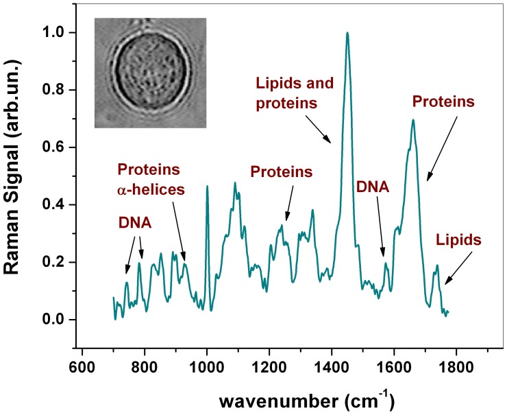
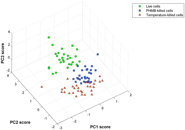
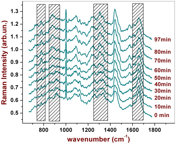
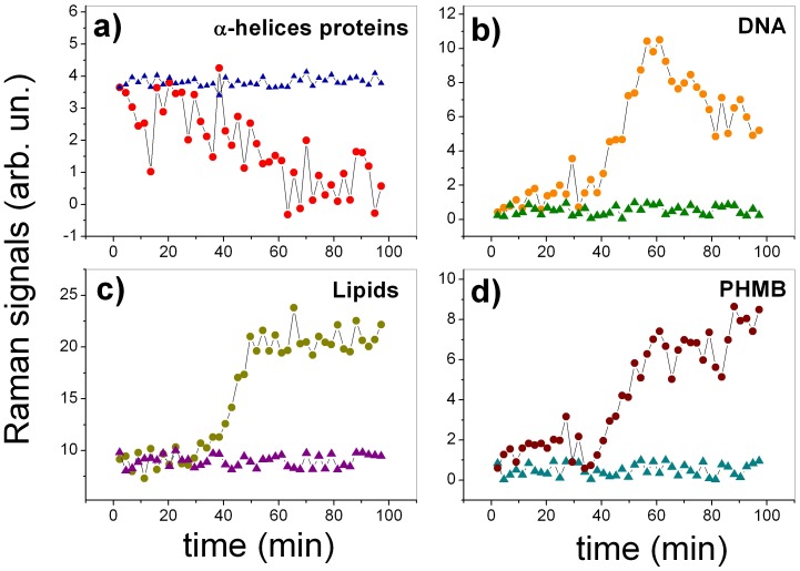
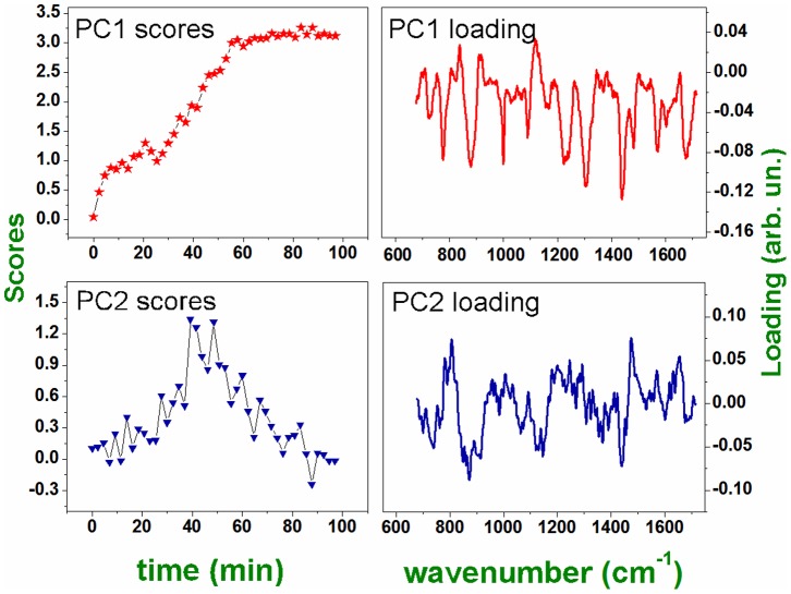
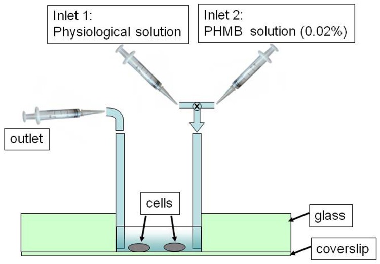
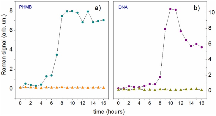
Acanthamoeba keratitis is a rare but serious corneal disease, often observed in contact lens wearers. Clinical treatment of infected patients frequently involves the use of polyhexamethylene biguanide (PHMB), a polymer used as a disinfectant and antiseptic, which is toxic also for the epithelial cells of the cornea. Prompt and effective diagnostic tools are hence highly desiderable for both starting early therapy and timely suspension of the treatment. In this work we use Raman microspectroscopy to analyse in vitro a single Acanthamoeba cell in cystic phase. In particular, we investigate the effect of PHMB at the single-cell level, providing useful information on both the underlying biochemical mechanism and the time frame for Acanthamoeba eradication in ocular infections. Furthermore, we demonstrate that Raman spectroscopy, in conjunction with standard multivariate analysis methods, allows discriminating between live and dead Acanthamoebas, which is fundamental to optimizing patients' treatment.
棘阿米巴角膜炎是一种罕见但严重的角膜疾病,常发生在隐形眼镜佩戴者中。受感染患者的临床治疗经常涉及使用聚六亚甲基双胍(PHMB),这是一种用作消毒剂和防腐剂的聚合物,对角膜的上皮细胞也有毒性。因此,对于早期开始治疗和及时停止治疗,都迫切需要快速有效的诊断工具。在这项工作中,我们使用拉曼微光谱技术分析处于囊包期的单个棘阿米巴细胞。特别是,我们研究了 PHMB 在单细胞水平上的作用,为眼部感染中棘阿米巴的消除的潜在生化机制和时间框架提供了有用的信息。此外,我们证明了拉曼光谱结合标准多元分析方法,可以区分死活棘阿米巴,这对于优化患者的治疗至关重要。