Mao Jianhua, Wang Dayan, Mataleena Parikka, He Bing, Niu Dadi, Katayama Kan, Xu Xiangjun, Ojala Juha Rm, Wang Wenjing, Shu Qiang, Du Lizhong, Liu Aimin, Pikkarainen Timo, Patrakka Jaakko, Tryggvason Karl
Department of Nephrology, The Children's Hospital of Zhejiang University School of Medicine, Hangzhou, PR China.
PLoS One. 2013 Aug 19;8(8):e72750. doi: 10.1371/journal.pone.0072750. eCollection 2013.
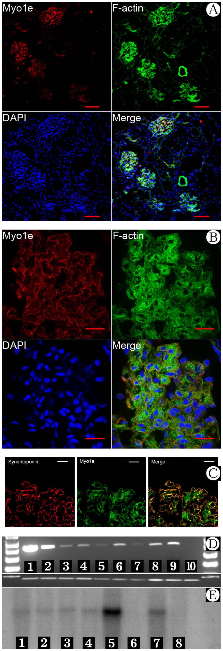
Podocytes serve as an important constituent of the glomerular filtration barrier. Recently, we and others identified Myo1e as a key molecular component of the podocyte cytoskeleton.
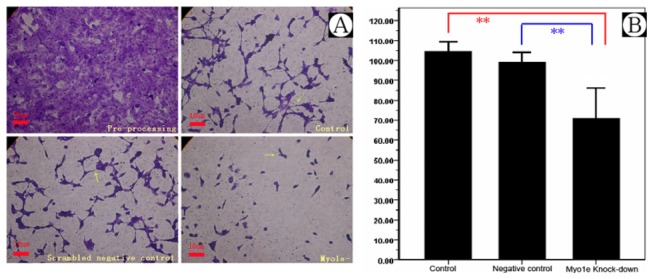
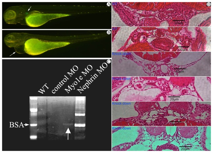
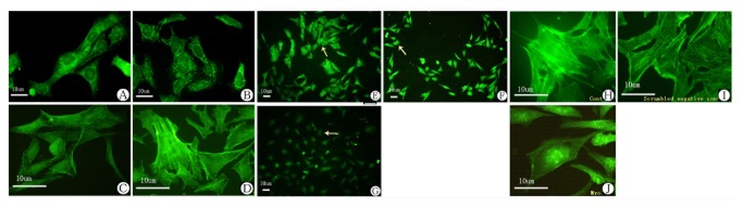
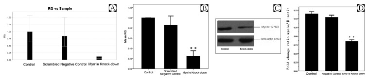
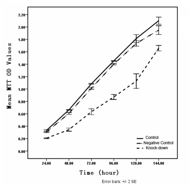
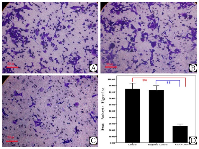
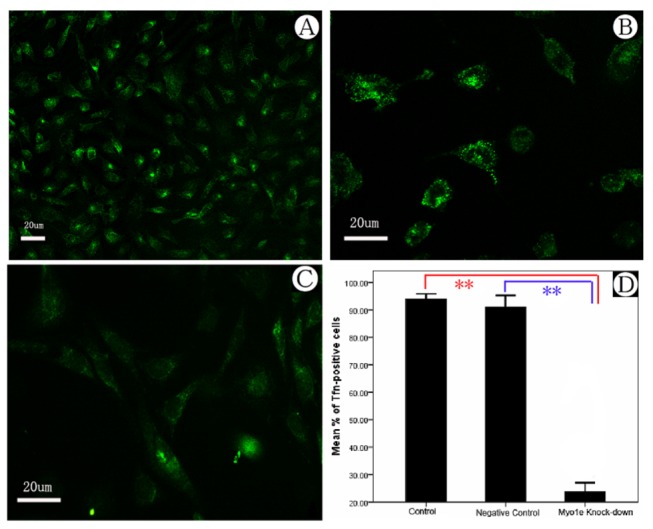
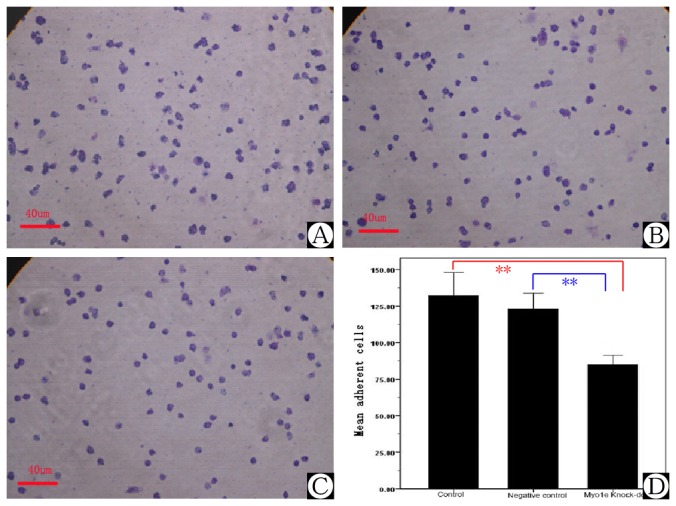
Myo1e mRNA and protein was expressed in human and mouse kidney sections as determined by Northern blot and reverse transcriptase PCR, and its expression was more evident in podocytes by immunofluorescence. By specific knock-down of MYO1E in zebrafish, the injected larvae exhibited pericardial edema and pronephric cysts, consistent with the appearance of protein in condensed incubation supernate. Furthermore, specific inhibition of Myo1e expression in a conditionally immortalized podocyte cell line induced morphological changes, actin cytoskeleton rearrangement, and dysfunction in cell proliferation, migration, endocytosis, and adhesion with the glomerular basement membrane.
Our results revealed that Myo1e is a key component contributing to the functional integrity of podocytes. Its impairment may cause actin cytoskeleton re-organization, alteration of cell shape, and membrane transport, and podocyte drop-out from the glomerular basement membrane, which might eventually lead to an impaired glomerular filtration barrier and proteinuria.
足细胞是肾小球滤过屏障的重要组成部分。最近,我们和其他研究人员确定Myo1e是足细胞细胞骨架的关键分子成分。
通过Northern印迹和逆转录酶PCR检测,Myo1e mRNA和蛋白在人和小鼠肾脏切片中表达,免疫荧光显示其在足细胞中的表达更明显。通过在斑马鱼中特异性敲低MYO1E,注射的幼虫出现心包水肿和前肾囊肿,这与浓缩孵育上清液中蛋白质的出现情况一致。此外,在条件永生化足细胞系中特异性抑制Myo1e表达会诱导形态变化、肌动蛋白细胞骨架重排以及细胞增殖、迁移、内吞作用和与肾小球基底膜粘附功能障碍。
我们的结果表明,Myo1e是维持足细胞功能完整性的关键成分。其受损可能导致肌动蛋白细胞骨架重组、细胞形状改变和膜转运,以及足细胞从肾小球基底膜脱落,最终可能导致肾小球滤过屏障受损和蛋白尿。