Department of Knowledge Engineering, Maastricht University, Maastricht, The Netherlands.
PLoS Comput Biol. 2013;9(8):e1003202. doi: 10.1371/journal.pcbi.1003202. Epub 2013 Aug 22.
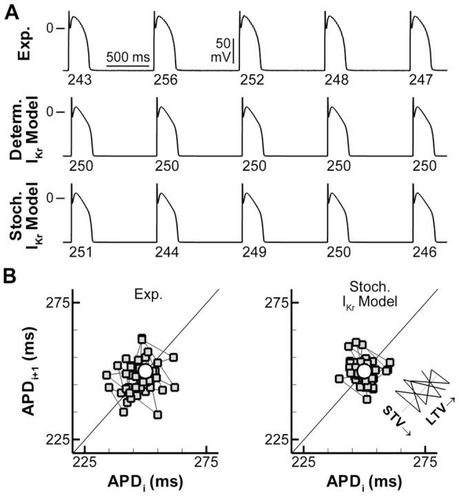
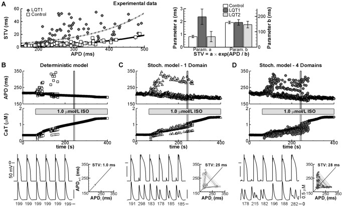
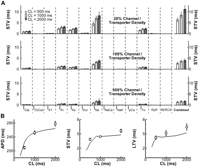
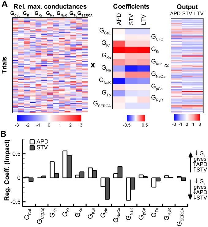
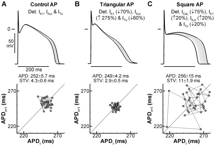
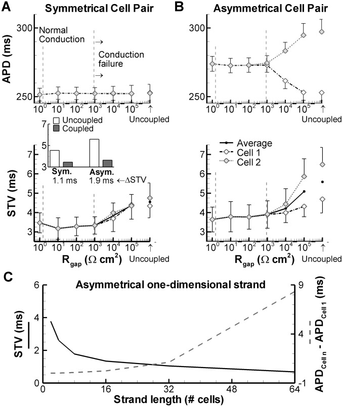
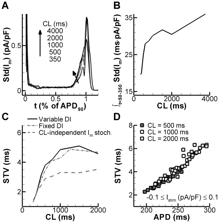
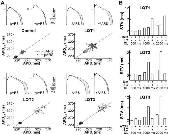
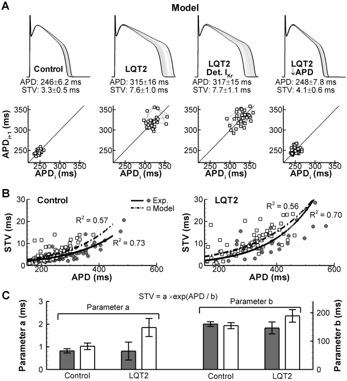
Beat-to-beat variability of repolarization duration (BVR) is an intrinsic characteristic of cardiac function and a better marker of proarrhythmia than repolarization prolongation alone. The ionic mechanisms underlying baseline BVR in physiological conditions, its rate dependence, and the factors contributing to increased BVR in pathologies remain incompletely understood. Here, we employed computer modeling to provide novel insights into the subcellular mechanisms of BVR under physiological conditions and during simulated drug-induced repolarization prolongation, mimicking long-QT syndromes type 1, 2, and 3. We developed stochastic implementations of 13 major ionic currents and fluxes in a model of canine ventricular-myocyte electrophysiology. Combined stochastic gating of these components resulted in short- and long-term variability, consistent with experimental data from isolated canine ventricular myocytes. The model indicated that the magnitude of stochastic fluctuations is rate dependent due to the rate dependence of action-potential (AP) duration (APD). This process (the "active" component) and the intrinsic nonlinear relationship between membrane current and APD ("intrinsic component") contribute to the rate dependence of BVR. We identified a major role in physiological BVR for stochastic gating of the persistent Na(+) current (INa) and rapidly activating delayed-rectifier K(+) current (IKr). Inhibition of IKr or augmentation of INa significantly increased BVR, whereas subsequent β-adrenergic receptor stimulation reduced it, similar to experimental findings in isolated myocytes. In contrast, β-adrenergic stimulation increased BVR in simulated long-QT syndrome type 1. In addition to stochastic channel gating, AP morphology, APD, and beat-to-beat variations in Ca(2+) were found to modulate single-cell BVR. Cell-to-cell coupling decreased BVR and this was more pronounced when a model cell with increased BVR was coupled to a model cell with normal BVR. In conclusion, our results provide new insights into the ionic mechanisms underlying BVR and suggest that BVR reflects multiple potentially proarrhythmic parameters, including increased ion-channel stochasticity, prolonged APD, and abnormal Ca(2+) handling.
心动周期复极时程变异(BVR)是心脏功能的固有特征,是比单纯复极延长更好的致心律失常标志物。生理条件下 BVR 的基础离子机制、其频率依赖性以及导致病变中 BVR 增加的因素仍不完全清楚。在这里,我们采用计算机建模,为生理条件下和模拟药物诱导复极延长下(模拟 1 型、2 型和 3 型长 QT 综合征)BVR 的亚细胞机制提供新的见解。我们在犬心室肌细胞电生理学模型中开发了 13 种主要离子电流和通量的随机实现。这些组件的随机门控组合导致了短期和长期的变异性,与从分离的犬心室肌细胞获得的实验数据一致。模型表明,由于动作电位(AP)时程(APD)的频率依赖性,随机波动的幅度是频率依赖性的。这个过程(“主动”成分)和膜电流与 APD 之间的固有非线性关系(“固有”成分)有助于 BVR 的频率依赖性。我们发现,在生理 BVR 中,持续钠电流(INa)和快速激活延迟整流钾电流(IKr)的随机门控起主要作用。IKr 抑制或 INa 增强显著增加了 BVR,而随后的β-肾上腺素能受体刺激降低了 BVR,这与分离肌细胞中的实验结果相似。相比之下,β-肾上腺素能刺激增加了模拟 1 型长 QT 综合征中的 BVR。除了随机通道门控外,AP 形态、APD 和 Ca2+的心动周期变化被发现调节单细胞 BVR。细胞间耦合降低了 BVR,当具有增加 BVR 的模型细胞与具有正常 BVR 的模型细胞耦合时,这种情况更为明显。总之,我们的结果提供了 BVR 基础离子机制的新见解,并表明 BVR 反映了多个潜在的致心律失常参数,包括增加的离子通道随机性、延长的 APD 和异常的 Ca2+处理。