• 文献检索
  • 文档翻译
  • 深度研究
  • 学术资讯
  • Suppr Zotero 插件Zotero 插件
  • 邀请有礼
  • 套餐&价格
  • 历史记录
应用&插件
Suppr Zotero 插件Zotero 插件浏览器插件Mac 客户端Windows 客户端微信小程序
定价
高级版会员购买积分包购买API积分包
服务
文献检索文档翻译深度研究API 文档MCP 服务
关于我们
关于 Suppr公司介绍联系我们用户协议隐私条款
关注我们

Suppr 超能文献

核心技术专利:CN118964589B侵权必究
粤ICP备2023148730 号-1Suppr @ 2026

文献检索

告别复杂PubMed语法,用中文像聊天一样搜索,搜遍4000万医学文献。AI智能推荐,让科研检索更轻松。

立即免费搜索

文件翻译

保留排版,准确专业,支持PDF/Word/PPT等文件格式,支持 12+语言互译。

免费翻译文档

深度研究

AI帮你快速写综述,25分钟生成高质量综述,智能提取关键信息,辅助科研写作。

立即免费体验

脑肿瘤的 7.0T MRI 增强前后的早期经验。

Early experience of pre- and post-contrast 7.0T MRI in brain tumors.

机构信息

Department of Neurosurgery, Seoul National University Hospital, Seoul, Korea.

出版信息

J Korean Med Sci. 2013 Sep;28(9):1362-72. doi: 10.3346/jkms.2013.28.9.1362. Epub 2013 Aug 28.

DOI:10.3346/jkms.2013.28.9.1362
PMID:24015044
原文链接:https://pmc.ncbi.nlm.nih.gov/articles/PMC3763113/
Abstract

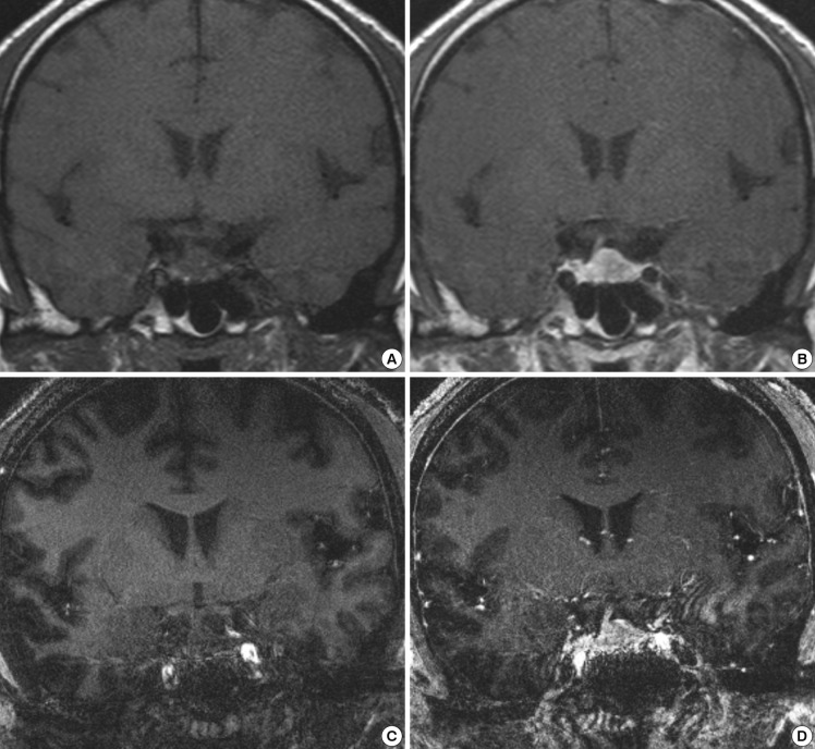
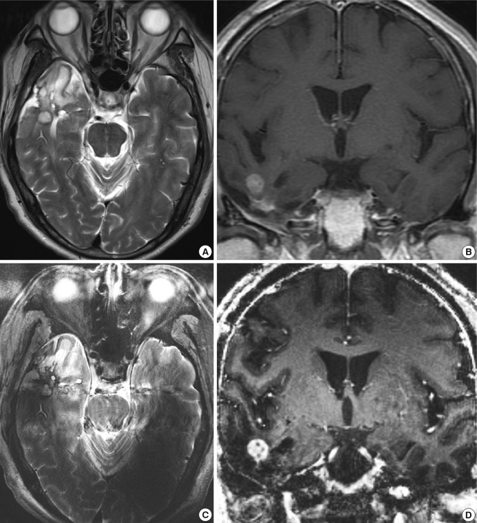
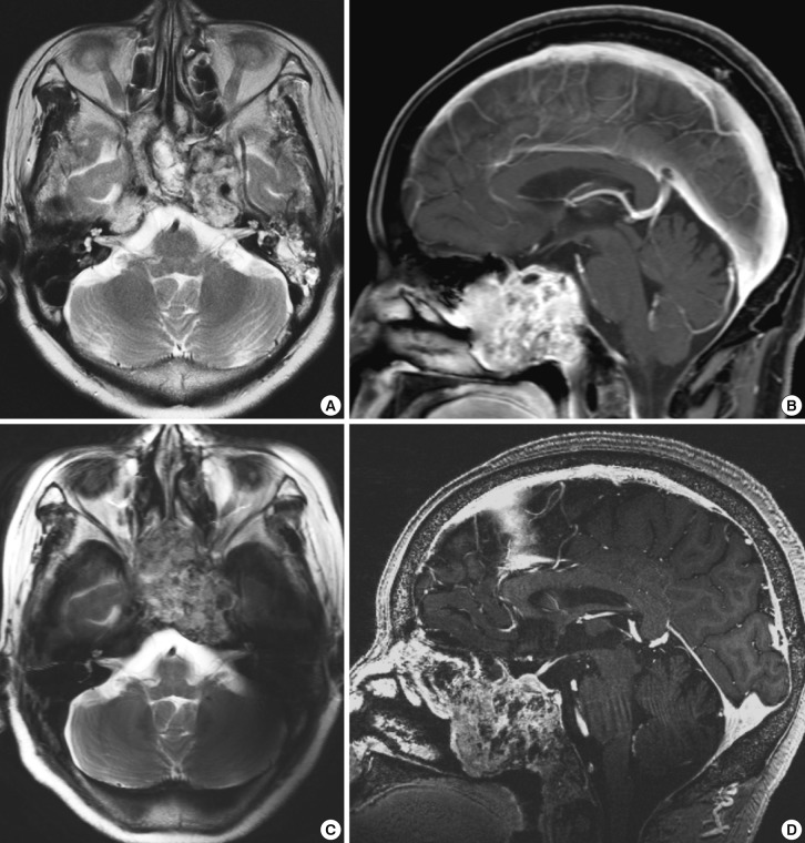
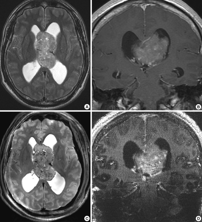
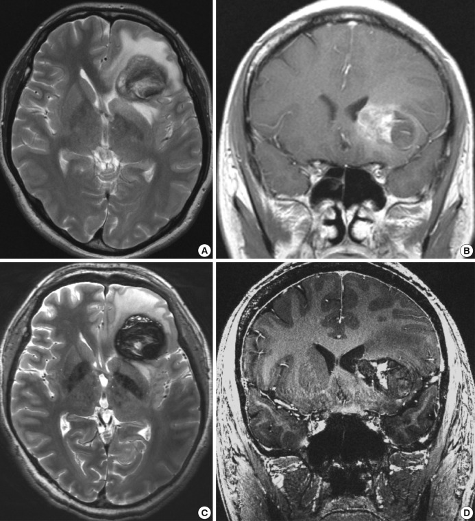
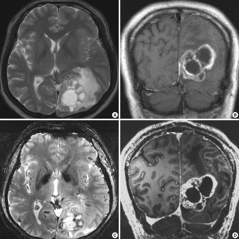
We investigated the safety and clinical applicability of 7.0 Tesla (T) brain magnetic resonance imaging (MRI) in patients with brain tumors. Twenty-four patients with intraaxial or extraaxial brain tumors were enrolled in this study. 7.0T MRIs of T2*-weighted axial and T1-weighted coronal or sagittal images were obtained and compared with 1.5T brain MRIs. The T2*-weighted images from 7.0T brain MRI revealed detailed microvasculature and the internal contents of supratentorial brain tumors better than that of 1.5T brain MRI. For brain tumors located in parasellar areas or areas adjacent to major cerebral vessels, flow-related artifacts were exaggerated in the 7.0T brain MRIs. For brain tumors adjacent to the skull base, susceptibility artifacts in the interfacing areas of the paranasal sinus and skull base hampered the aquisition of detailed images and information on brain tumors in the 7.0T brain MRIs. This study shows that 7.0T brain MRI can provide detailed information on the intratumoral components and margins in supratentorial brain tumors. Further studies are needed to develop refined MRI protocols for better images of brain tumors located in the skull base, parasellar, and adjacent major cerebrovascular structures.

摘要

我们研究了 7.0 特斯拉(T)脑部磁共振成像(MRI)在脑肿瘤患者中的安全性和临床适用性。本研究纳入了 24 例脑内或脑外肿瘤患者。获取了 T2*-加权轴位和 T1 加权冠状或矢状位 7.0T MRI,并与 1.5T 脑部 MRI 进行了比较。7.0T 脑部 MRI 的 T2*-加权图像比 1.5T 脑部 MRI 更能清晰显示脑肿瘤的微血管和内部结构。对于鞍旁区或毗邻主要脑血管的脑肿瘤,7.0T 脑部 MRI 中夸大了与血流相关的伪影。对于毗邻颅底的脑肿瘤,副鼻窦和颅底交界区的磁敏感伪影妨碍了详细的肿瘤图像和信息获取。本研究表明,7.0T 脑部 MRI 可以提供脑内肿瘤成分和边界的详细信息。需要进一步研究以开发更精细的 MRI 方案,以更好地显示颅底、鞍旁和毗邻主要脑血管结构的脑肿瘤。

https://cdn.ncbi.nlm.nih.gov/pmc/blobs/3f1a/3763113/c6ae31bbeec0/jkms-28-1362-g006.jpg
https://cdn.ncbi.nlm.nih.gov/pmc/blobs/3f1a/3763113/32bb4583adec/jkms-28-1362-g001.jpg
https://cdn.ncbi.nlm.nih.gov/pmc/blobs/3f1a/3763113/6b25c1950bc1/jkms-28-1362-g002.jpg
https://cdn.ncbi.nlm.nih.gov/pmc/blobs/3f1a/3763113/bfe0afde0883/jkms-28-1362-g003.jpg
https://cdn.ncbi.nlm.nih.gov/pmc/blobs/3f1a/3763113/4a08ebf1b871/jkms-28-1362-g004.jpg
https://cdn.ncbi.nlm.nih.gov/pmc/blobs/3f1a/3763113/ad85c2ac3cf7/jkms-28-1362-g005.jpg
https://cdn.ncbi.nlm.nih.gov/pmc/blobs/3f1a/3763113/c6ae31bbeec0/jkms-28-1362-g006.jpg

相似文献

1
Early experience of pre- and post-contrast 7.0T MRI in brain tumors.脑肿瘤的 7.0T MRI 增强前后的早期经验。
J Korean Med Sci. 2013 Sep;28(9):1362-72. doi: 10.3346/jkms.2013.28.9.1362. Epub 2013 Aug 28.
2
A case report of preoperative and postoperative 7.0T brain MRI in a patient with a small cell glioblastoma.一例小细胞胶质母细胞瘤患者术前及术后7.0T脑部磁共振成像的病例报告。
J Korean Med Sci. 2014 Jul;29(7):1012-7. doi: 10.3346/jkms.2014.29.7.1012. Epub 2014 Jul 11.
3
Magnetic resonance imaging of hyaline cartilage defects at 1.5T and 3.0T: comparison of medium T2-weighted fast spin echo, T1-weighted two-dimensional and three-dimensional gradient echo pulse sequences.1.5T和3.0T下透明软骨缺损的磁共振成像:中等T2加权快速自旋回波、T1加权二维和三维梯度回波脉冲序列的比较
Acta Radiol. 2005 Feb;46(1):67-73. doi: 10.1080/02841850510012625.
4
Water excitation MPRAGE: an alternative sequence for postcontrast imaging of the abdomen in noncooperative patients at 1.5 Tesla and 3.0 Tesla MRI.水激发MPRAGE序列:一种用于1.5特斯拉和3.0特斯拉MRI下非合作患者腹部增强成像的替代序列。
J Magn Reson Imaging. 2008 May;27(5):1146-54. doi: 10.1002/jmri.21346.
5
Magnetic resonance imaging of the cranial nerves in the posterior fossa: a comparative study of t2-weighted spin-echo sequences at 1.5 and 3.0 tesla.后颅窝颅神经的磁共振成像:1.5特斯拉和3.0特斯拉T2加权自旋回波序列的对比研究
Acta Radiol. 2008 Apr;49(3):358-63. doi: 10.1080/02841850701824127.
6
Effect of superparamagnetic iron oxide on tumor-to-liver contrast at T2*-weighted gradient-echo MRI: comparison between 3.0T and 1.5T MR systems.超顺磁性氧化铁对T2*加权梯度回波磁共振成像中肿瘤与肝脏对比度的影响:3.0T和1.5T磁共振系统的比较
J Magn Reson Imaging. 2009 Mar;29(3):595-600. doi: 10.1002/jmri.21384.
7
Diagnostic comparison of 1.5 Tesla and 3.0 Tesla preoperative MRI of the wrist in patients with ulnar-sided wrist pain.尺侧腕部疼痛患者术前1.5特斯拉和3.0特斯拉腕部MRI的诊断比较
J Hand Surg Am. 2008 Sep;33(7):1153-9. doi: 10.1016/j.jhsa.2008.02.028.
8
Fluid attenuated inversion recovery (FLAIR) imaging of the normal brain: comparisons between under the conditions of 3.0 Tesla and 1.5 Tesla.正常脑的液体衰减反转恢复(FLAIR)成像:3.0 特斯拉与 1.5 特斯拉条件下的比较。
Korean J Radiol. 2010 Jan-Feb;11(1):19-24. doi: 10.3348/kjr.2010.11.1.19. Epub 2009 Dec 28.
9
Quantitative and qualitative comparison of 3.0T and 1.5T MR imaging of the liver in patients with diffuse parenchymal liver disease.弥漫性实质性肝病患者肝脏 3.0T 与 1.5T MR 成像的定量和定性比较。
Eur J Radiol. 2009 Nov;72(2):314-20. doi: 10.1016/j.ejrad.2008.07.027. Epub 2008 Sep 13.
10
Fast fluid-attenuated inversion-recovery (FLAIR) MRI in the assessment of intraaxial brain tumors.快速液体衰减反转恢复(FLAIR)磁共振成像在评估脑内肿瘤中的应用
J Magn Reson Imaging. 1998 Jul-Aug;8(4):789-98. doi: 10.1002/jmri.1880080407.

引用本文的文献

1
Radiomics-driven neuro-fuzzy framework for rule generation to enhance explainability in MRI-based brain tumor segmentation.基于放射组学的神经模糊框架用于生成规则以增强基于MRI的脑肿瘤分割中的可解释性。
Front Neuroinform. 2025 Apr 17;19:1550432. doi: 10.3389/fninf.2025.1550432. eCollection 2025.
2
Use of a Commercial 7-T MRI Scanner for Clinical Brain Imaging: Indications, Protocols, Challenges, and Solutions-A Single-Center Experience.商用 7T MRI 扫描仪在临床脑部成像中的应用:单中心经验分享——适应证、方案、挑战及解决方案。
AJR Am J Roentgenol. 2023 Dec;221(6):788-804. doi: 10.2214/AJR.23.29342. Epub 2023 Jun 28.
3

本文引用的文献

1
Clinical applications of 7 T MRI in the brain.7T MRI 在脑部的临床应用。
Eur J Radiol. 2013 May;82(5):708-18. doi: 10.1016/j.ejrad.2011.07.007. Epub 2011 Sep 19.
2
Effect of the extracranial deep brain stimulation lead on radiofrequency heating at 9.4 Tesla (400.2 MHz).体外深脑刺激导线对 9.4T(400.2MHz)射频加热的影响。
J Magn Reson Imaging. 2010 Sep;32(3):600-7. doi: 10.1002/jmri.22292.
3
Imaging of adult astrocytic brain tumours with 7 T MRI: preliminary results.7T MRI 对成人星形细胞瘤的成像:初步结果。
MRI with ultrahigh field strength and high-performance gradients: challenges and opportunities for clinical neuroimaging at 7 T and beyond.
具有超高场强和高性能梯度的磁共振成像:7T及更高场强下临床神经成像面临的挑战与机遇
Eur Radiol Exp. 2021 Aug 26;5(1):35. doi: 10.1186/s41747-021-00216-2.
4
Emerging Use of Ultra-High-Field 7T MRI in the Study of Intracranial Vascularity: State of the Field and Future Directions.超高场 7T MRI 在颅内血管研究中的新兴应用:现状与未来方向。
AJNR Am J Neuroradiol. 2020 Jan;41(1):2-9. doi: 10.3174/ajnr.A6344. Epub 2019 Dec 26.
5
First Application of 7-T Magnetic Resonance Imaging in Endoscopic Endonasal Surgery of Skull Base Tumors.7-T磁共振成像在颅底肿瘤鼻内镜鼻内手术中的首次应用。
World Neurosurg. 2017 Jul;103:600-610. doi: 10.1016/j.wneu.2017.03.088. Epub 2017 Mar 27.
6
Comparison of 3 and 7 Tesla Magnetic Resonance Imaging of Obstructive Hydrocephalus Caused by Tectal Glioma.顶盖胶质瘤所致梗阻性脑积水的3特斯拉与7特斯拉磁共振成像比较
Brain Tumor Res Treat. 2016 Oct;4(2):150-154. doi: 10.14791/btrt.2016.4.2.150. Epub 2016 Oct 31.
7
A critical reflection on the technological development of deep brain stimulation (DBS).深度脑刺激 (DBS) 技术发展的批判性反思。
Front Hum Neurosci. 2014 Sep 17;8:730. doi: 10.3389/fnhum.2014.00730. eCollection 2014.
8
The evolving role of neurological imaging in neuro-oncology.神经影像学在神经肿瘤学中不断演变的作用。
J Neurooncol. 2014 Sep;119(3):491-502. doi: 10.1007/s11060-014-1505-3. Epub 2014 Aug 1.
9
A case report of preoperative and postoperative 7.0T brain MRI in a patient with a small cell glioblastoma.一例小细胞胶质母细胞瘤患者术前及术后7.0T脑部磁共振成像的病例报告。
J Korean Med Sci. 2014 Jul;29(7):1012-7. doi: 10.3346/jkms.2014.29.7.1012. Epub 2014 Jul 11.
Eur Radiol. 2010 Mar;20(3):704-13. doi: 10.1007/s00330-009-1592-2. Epub 2009 Sep 18.
4
GRAPPA-based susceptibility-weighted imaging of normal volunteers and patients with brain tumor at 7 T.基于GRAPPA的7T场强下正常志愿者和脑肿瘤患者的 susceptibility加权成像 。 (注:susceptibility可能存在专业术语翻译错误,可根据具体医学内容准确翻译,比如“磁敏感”等 )
Magn Reson Imaging. 2009 May;27(4):480-8. doi: 10.1016/j.mri.2008.08.003. Epub 2008 Sep 26.
5
Observation of the lenticulostriate arteries in the human brain in vivo using 7.0T MR angiography.使用7.0T磁共振血管造影术对人脑豆纹动脉进行活体观察。
Stroke. 2008 May;39(5):1604-6. doi: 10.1161/STROKEAHA.107.508002. Epub 2008 Mar 13.
6
High-field, high-resolution, susceptibility-weighted magnetic resonance imaging: improved image quality by addition of contrast agent and higher field strength in patients with brain tumors.高场、高分辨率、磁化率加权磁共振成像:通过添加造影剂和提高场强改善脑肿瘤患者的图像质量
Neuroradiology. 2008 Jan;50(1):9-16. doi: 10.1007/s00234-007-0298-x. Epub 2007 Sep 18.
7
The 2007 WHO classification of tumours of the central nervous system.2007年世界卫生组织中枢神经系统肿瘤分类
Acta Neuropathol. 2007 Aug;114(2):97-109. doi: 10.1007/s00401-007-0243-4. Epub 2007 Jul 6.
8
Clinical magnetic resonance imaging of brain tumors at ultrahigh field: a state-of-the-art review.超高场强下脑肿瘤的临床磁共振成像:最新综述
Top Magn Reson Imaging. 2006 Apr;17(2):53-61. doi: 10.1097/RMR.0b013e3180300404.
9
Limits of 8-Tesla magnetic resonance imaging spatial resolution of the deoxygenated cerebral microvasculature.8特斯拉磁共振成像对脱氧脑微血管系统空间分辨率的局限性。
J Magn Reson Imaging. 2004 Mar;19(3):303-7. doi: 10.1002/jmri.20006.
10
Controlled E-field gradient coils.可控电场梯度线圈。
MAGMA. 2003 Nov;16(3):113-20. doi: 10.1007/s10334-003-0015-7. Epub 2003 Oct 31.