First Department of Critical Care and Pulmonary Services, Evangelismos Hospital, University of Athens Medical School, Athens, Greece ; G. P. Livanos and M. Simou Laboratories, Evangelismos Hospital, University of Athens Medical School, Athens, Greece.
Pulm Circ. 2013 Apr;3(2):419-25. doi: 10.4103/2045-8932.113189.
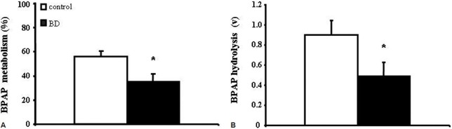
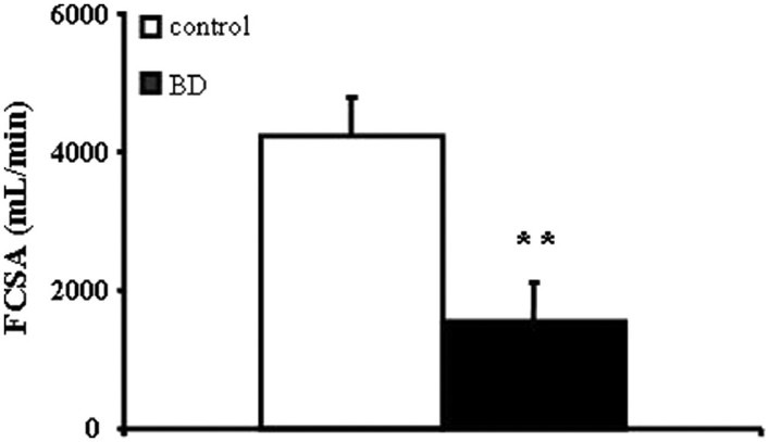
Pulmonary endothelium is a major metabolic organ affecting pulmonary and systemic vascular homeostasis. Brain death (BD)-induced physiologic and metabolic derangements in donors' lungs, in the absence of overt lung pathology, may cause pulmonary dysfunction and compromise post-transplant graft function. To explore the impact of BD on pulmonary endothelium, we estimated pulmonary capillary endothelium-bound (PCEB)-angiotensin converting enzyme (ACE) activity, a direct and quantifiable index of pulmonary endothelial function, in eight brain-dead patients and ten brain-injured mechanically ventilated controls. No subject suffered from acute lung injury or any other overt lung pathology. Applying indicator-dilution type techniques, we measured single-pass transpulmonary percent metabolism (%M) and hydrolysis (v) of the synthetic, biologically inactive, and highly specific for ACE substrate (3)H-benzoyl-Phe-Ala-Pro, under first order reaction conditions, and calculated lung functional capillary surface area (FCSA). Substrate %M (35 ± 6.8%) and v (0.49 ± 0.13) in BD patients were decreased as compared to controls (55.9 ± 4.9, P = 0.033 and 0.9 ± 0.15, P = 0.033, respectively), denoting decreased pulmonary endothelial enzyme activity at the capillary level; FCSA, a reflection of endothelial enzyme activity per vascular bed, was also decreased (BD patients: 1,563 ± 562 mL/min vs 4,235 ± 559 in controls; P = 0.003). We conclude that BD is associated with subtle pulmonary endothelial injury, expressed by decreased PCEB-ACE activity. The applied indicator-dilution type technique provides direct and quantifiable indices of pulmonary endothelial function at the bedside that may reveal the existence of preclinical lung pathology in potential lung donors.
肺内皮细胞是影响肺和全身血管内稳态的主要代谢器官。在没有明显肺部病理的情况下,脑死亡(BD)引起供体肺部的生理和代谢紊乱,可能导致肺功能障碍,并影响移植后移植物的功能。为了探讨 BD 对肺内皮细胞的影响,我们评估了 8 例脑死亡患者和 10 例脑损伤机械通气对照者肺毛细血管内皮结合(PCEB)-血管紧张素转换酶(ACE)活性,这是一种直接和可量化的肺内皮功能指标。没有受试者患有急性肺损伤或任何其他明显的肺部疾病。应用指示剂稀释型技术,我们在第一级反应条件下测量了合成的、生物上无活性的、高度特异的 ACE 底物(3)H-苯甲酰-Phe-Ala-Pro 的单pass 经肺代谢百分比(%M)和水解(v),并计算了肺功能性毛细血管表面积(FCSA)。与对照组相比,BD 患者的底物%M(35 ± 6.8%)和 v(0.49 ± 0.13)降低,分别为(P = 0.033 和 P = 0.033),这表示毛细血管水平肺内皮酶活性降低;反映每个血管床内皮酶活性的 FCSA 也降低(BD 患者:1563 ± 562 mL/min 与对照组的 4235 ± 559;P = 0.003)。我们的结论是,BD 与肺内皮的细微损伤有关,表现为 PCEB-ACE 活性降低。应用的指示剂稀释型技术可提供床边直接和可量化的肺内皮功能指标,这可能揭示潜在肺供体临床前肺病理的存在。