Reddy Rahul K, Lalezary Maziar, Kim Stephen J, Kammer Jeffrey A, Kuchtey Rachel W, Cherney Edward F, Recchia Franco M, Joos Karen M, Agarwal Anita, Law Janice C
Department of Ophthalmology, Vanderbilt University School of Medicine, Nashville, TN, USA.
Clin Ophthalmol. 2013;7:1761-9. doi: 10.2147/OPTH.S49375. Epub 2013 Sep 2.
The purpose of this paper is to report the 3-month findings of the Prospective Retinal and Optic Nerve Vitrectomy Evaluation (PROVE) study.
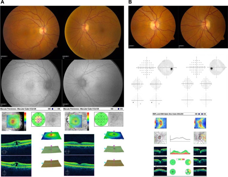
Eighty eyes of 40 participants undergoing vitrectomy were enrolled. Participants underwent baseline evaluation of the study (surgical) and fellow (control) eye that included: intraocular pressure, central corneal thickness, gonioscopy, cup-to-disc ratio measurement, color fundus and optic disc photography, automated perimetry, and optical coherence tomography of the macula and optic nerve. Evaluation was repeated at 3 months. Main outcome measures were changes in macula and retinal nerve fiber layer (RNFL) thickness and intraocular pressure.
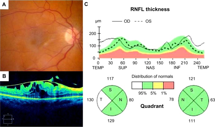
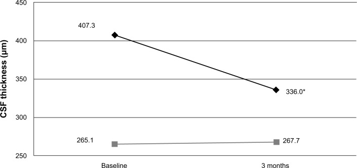
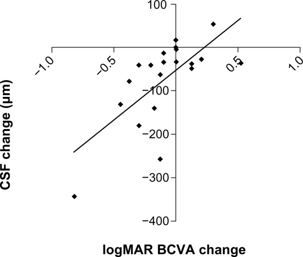
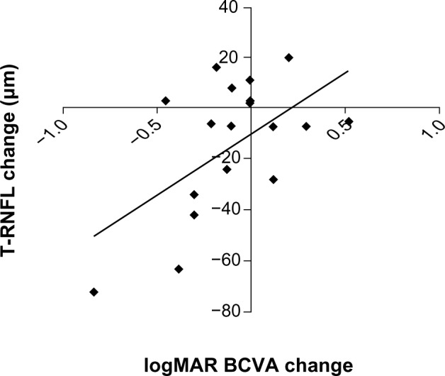
All participants completed follow-up. Mean cup-to-disc ratio of study and fellow eyes at baseline was 0.43 ± 0.2 and 0.46 ± 0.2, respectively, and 13% of participants had undiagnosed narrow angles. There was no significant change in intraocular pressure, cup-to-disc ratio, or pattern standard deviation in study eyes compared with baseline or fellow eyes at 3 months. Vision improved in all study eyes at 3 months compared with baseline (P = 0.013), but remained significantly worse than fellow eyes (P < 0.001). Central subfield and temporal peripapillary RNFL thickness were significantly greater in eyes with epiretinal membrane (P < 0.05), and resolution after surgery correlated with visual improvement (P < 0.05).
The 3-month results do not indicate any increased risk for open-angle glaucoma but suggest that a relatively high percentage of eyes may be at risk of angle closure glaucoma. Temporal RNFL thickness and central subfield were increased in eyes with epiretinal membrane, and resolution correlated with degree of visual recovery.
本文旨在报告前瞻性视网膜和视神经玻璃体切除术评估(PROVE)研究的3个月结果。
纳入40名接受玻璃体切除术患者的80只眼睛。参与者对研究眼(手术眼)和对侧眼(对照眼)进行基线评估,包括:眼压、中央角膜厚度、前房角镜检查、杯盘比测量、彩色眼底和视盘照相、自动视野检查以及黄斑和视神经的光学相干断层扫描。在3个月时重复评估。主要观察指标为黄斑和视网膜神经纤维层(RNFL)厚度以及眼压的变化。
所有参与者均完成随访。研究眼和对侧眼在基线时的平均杯盘比分别为0.43±0.2和0.46±0.2,13%的参与者存在未诊断出的窄房角。与基线或3个月时的对侧眼相比,研究眼的眼压、杯盘比或模式标准差无显著变化。与基线相比,所有研究眼在3个月时视力均有改善(P = 0.013),但仍显著低于对侧眼(P < 0.001)。视网膜前膜患者的中央子野和视乳头周围颞侧RNFL厚度显著更大(P < 0.05),术后的恢复情况与视力改善相关(P < 0.05)。
3个月的结果未表明开角型青光眼风险增加,但提示相当比例的眼睛可能有闭角型青光眼风险。视网膜前膜患者的颞侧RNFL厚度和中央子野增加,恢复情况与视力恢复程度相关。